• Biology

    Biology

    The atom, biological macromolecules (carbohydrates, lípids, proteins, nucleic acids), cell biology, cancer

  • Drugs

    Drugs

    Drug development, pharmacodynamics, pharmacokinetics, toxicology

  • Nanomedicine

    Nanomedicine

    Introduction to nanotechnology, diagnostic devices, drug delivery, regenerative medicine

  • Miscellaneous

    Miscellaneous

    A more in-depth explanation of topics previously mentioned in the other sections

4.3.2. Cell respiration II: Oxidative phosphorylation

0 comments
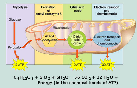
Most of the ATP originated during the aerobic catabolism of glucose does not come from the two previous routes (Glycolysis and Krebs cycle), but rather from the movement of electrons through a series of electron carriers that undergo redox reactions, which causes the accumulation of hydrogen ions in the mitochondrial matrix.

As a consequence, a concentration gradient is formed in which hydrogen ions diffuse out of the mitochondrial matrix by passing through it with the help of the complex called ATP synthase[1]. The current of hydrogen ions promotes the catalytic action of ATP synthase, which phosphorylates ADP, producing ATP.


Electron transport chain

The electron transport chain is the last component of aerobic respiration and the last stage of glucose metabolism that uses atmospheric oxygen.
This oxygen is used as a final receptor for the electrons that have been removed from the intermediate compounds in glucose catabolism.

The electron transport chain consists of four large multiprotein complexes (called complexes I-IV) embedded in the inner mitochondrial membrane and two small electron transporters that move the electrons between them.

Electrons participate in a series of redox reactions in which free energy is used to shuttle hydrogen ions across the mitochondrial membrane using the ATP synthase enzyme. This process contributes to the gradient used in chemiosmosis (a mechanism based on the diffusion of ions across a membrane) that produces 90% of ATP created during aerobic glucose metabolism.
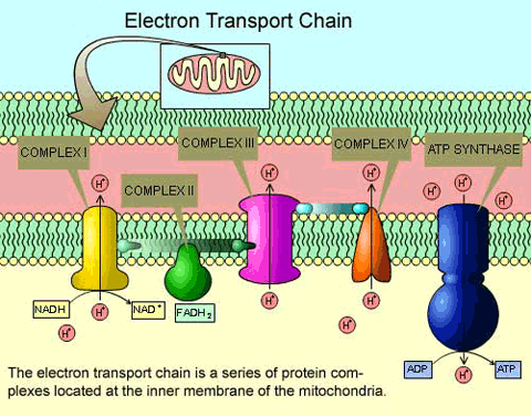
Elements of the electron transport chain

The electrons that take part in the transport chain lose their energy gradually, so to complete it, the contribution of high-energy electrons from the NADH or FADH2 compounds is necessary (generated in the previous process (Krebs cycle).

At the end of this metabolic pathway, electrons reduce oxygen molecules to oxygen ions and the extra electrons of these ions attract hydrogen ions (protons) from the surrounding environment, resulting in the release of water molecules and ATP as the final products of the electron transport chain.

The number of molecules created varies depending on a number of factors such as the number of hydrogen ions that the complexes of the electron chain pump across the mitochondrial membrane, the transport of electrons or the use of intermediate compounds produced in these routes for other purposes.


Metabolism without oxygen

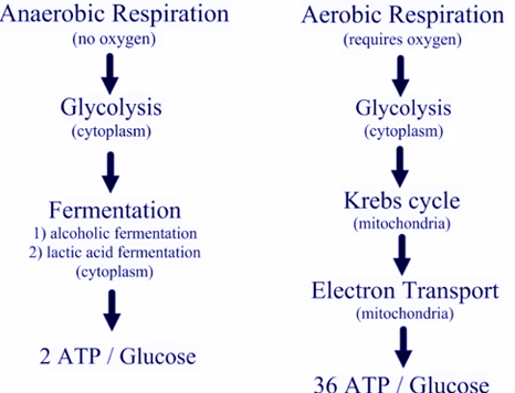
In aerobic respiration, the final receptor of electrons is the oxygen molecule (O2) and ATP is produced with the assistance of high-energy electrons transported to the transport chain by the molecules NADH or FADH2.
If aerobic respiration does not take place, the NADH compound must be re-oxidased to NAD+ so that it can be reused as an electron carrier, and in this way the glycolytic pathway continues.
Living organisms employ two different mechanisms to achieve this:
      They can use an organic molecule as the final acceptor of electrons to regenerate NAD+ from NADH in a process called fermentation.
One of the most well known fermentation processes is lactic acid fermentation, used by red blood cells in skeletal muscles of mammals when they lack enough oxygen to continue aerobic respiration.
Lactic acid fermentation
      The second option is to use an inorganic molecule instead.
In both, organisms transform energy to use it in the absence of oxygen and they are known as anaerobic cellular respiration.
Aerobic and anaerobic processes


Regulation of cellular respiration

Cellular respiration must be regulated in order to supply the required amounts of energy at any time in the form of ATP.
The cell must regulate, therefore, its metabolism and for that have a broad variety of mechanisms.
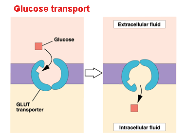
For instance, glucose entering the cell via the plasma membrane is controlled by transport proteins (GLUT proteins). But most of the control of the respiratory process is performed by specific enzymes that act on each route.
Cellular uptake of glucose
These enzymes react to the available levels of the nucleosides ATP, ADP, AMP, NAD+ and FAD, which, in turn, increase or decrease enzyme activity on the routes where they participate.

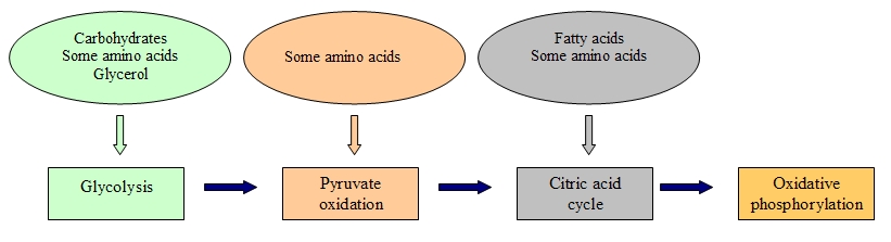
We have seen that glucose metabolism is responsible for providing energy to living cells. Yet, living beings consume a broad range of nutrients other than glucose in their diets. Hence, how do these foods become ATP in our cells?
Eventually the catabolic pathways of lipids, proteins and carbohydrates are connected with glycolysis and citric acid cycle pathways.
These pathways are not closed cycles, but many of their substrates, intermediate and final products are used in other routes.
Connection of carbohydrates, proteins and lipids to glucose metabolism


[1] It is a transmembrane protein complex (enzyme) that catalyses ATP synthesis by the supplied energy from a proton flow (H+) and by adding a phosphate group to ADP.  

Sources: OpenStax College, Biology. OpenStax College. 30 May 2013.
              https://chelseaharripersad.wordpress.com/2013/04/13/the-electron-transport-chain/
              http://healthylifemed.com/aerobic-vs-anaerobic-respiration/
              https://adapaproject.org/bbk_temp/tiki-index.php?page=Leaf%3A+Why+do+cells+need+fermentation+to+continue+glycolysis%3F
              http://163.178.103.176/Fisiologia/general/dinamica/FG05_17a.jpg


Read more
4.3.1. Cell respiration I: Glycolysis. Krebs cycle

0 comments
The energy that enters into our organisms as food is transformed so that cells can use it and perform their functions.
Thus, through a set of metabolic pathways (collectively called cell respiration), the energy of glucose bonds is extracted and turned into an energy form that all living organisms can use.
Metabolic pathways of cell respiration

Energy in living beings

A living cell does not collect excessive amounts of free energy because it could damage and subsequently destroy it. Hence, cells must store that energy in a safe way and free it only when the need arises, which is achieved by the adenosine triphosphate compound (ATP), which is considered the ‘energy currency’ of cells.

ATP allows the cell to store energy for a short time and to transport it within the cell to facilitate endergonic chemical reactions.
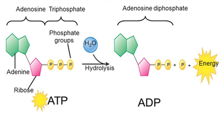
ATP’s structure is made up of an adenosine monophosphate molecule (AMP[1]), which consists of an adenine molecule attached to a ribose molecule and one phosphate group. The addition of a second phosphate group produces adenosine diphosphate (ADP) and the addition of a third phosphate group forms finally adenosine triphosphate (ATP).

Life to accomplish its processes and obtain energy, it breaks down ATP into ADP[2] constantly, through the hydrolysis ATP reaction, producing, at the same time, an inorganic phosphate ion:

ATP + H2O ADP + inorganic phosphate (Pi)
ATP hydrolysis

The water molecule that takes part in this reaction is split into a hydrogen atom and a hydroxyl group. This water is regenerated when a third phosphate is added to the ADP molecule, which reconstitutes, in turn, ATP. This mechanism works, therefore, like a rechargeable battery.

In practically all organisms, the required energy comes from glucose metabolism. This way, ATP is directly connected to the set of exergonic pathways of the catabolism of glucose and the large number of endergonic pathways that supply energy to cells.

The two processes of ATP regeneration used in combination with glucose catabolism are substrate phosphorylation[3] and oxidative phosphorylation.
In the first mechanism, ATP is produced from ADP and a phosphate group from a reactive.
But most of the generated ATP (90%) during the glucose catabolism is derived from a much more complex process, which ocurrs in the mitochondrion: chemiosmosis. During chemiosmosis in which a proton gradient is involved across the mitochondrial membrane.
The production of ATP using this mechanism is called oxidative phosphorylation because of the involvement of oxygen in it.


Glycolysis

Glycolysis was probably one of the first metabolic pathways used during the evolution of living beings and it is used by nearly all of them.
Glycolysis is the first point of glucose breakdown for the extraction of energy during the cellular metabolism. It does not require oxygen; it is thus an anaerobic mechanism.
Phases of glycolysis pathway
This process is composed of two steps:
      The six carbon atom ring of glucose molecule is divided into two sugar molecules (called pyruvate) with three carbon atoms each one. ATP is needed to cause this separation.
      ATP and high energy electrons are extracted from hydrogen atoms, and are attached to NAD+ compounds (oxidised form of the molecule NAD[4]).
The first stage invests two ATP molecules, while the second stage produces four ATP molecules through the substrate phosphorylation mechanism.
The final outcome is a net gain of two ATP molecules and two NADH[5] for the cell.
In the event that the cell cannot catalyse the pyruvate molecules, it only gets two ATP molecules from a glucose molecule.


Oxidation of pyruvate and the Citric Acid Cycle (Krebs cycle)

If there is oxygen available, aerobic respiration then takes place following the glycolysis process.
Pyruvate molecules originated at the end of that metabolic pathway are transported into mitochondria where cell respiration takes place. There, pyruvate is transformed into an acetyl group, which is collected and activated by a carrier called coenzyme A (CoA)..
The resultant compound, named acetyl coenzime A, is made up of vitamin B5 (pantothenic acid). This compound is used in a wide range of ways by cells, but its principal function is to distribute the acetyl group that comes from the pyruvate to the following stage in glucose metabolism (the citric acid cycle).
Krebs cycle scheme
The conversion of pyruvate to an acetyl group removes a CO2 molecule and two high energy electrons. This step occurs twice, so that the CO2 molecule (2CO2) holds two of the six carbon atoms from the initial molecule of glucose. While the NAD+ molecule picks up the electrons forming NADH that conducts those electrons to later pathways in the production of ATP.

At this point, the glucose molecule that entered the cellular respiration mechanism has been completely oxidised and its potential energy has been transferred to electron transporters or has been used to synthesise a few ATP molecules.

Next, the citric acid cycle also begins in the mitochondrial matrix.
Unlike glycolysis, the cytric acid cycle is a closed cycle, in which the last step regenerates the compound used in the first stage.
The cytric acid cycle is an eight step cycle that comprises a series of oxidation-reduction (redox), dehydration, hydration and decarboxylation reactions that produce: two CO2 molecules, one GTP[6]/ATP molecule and the reduced forms of three NADH molecules and one FADH2[7] molecule.

This cycle is considered an aerobic route because these two last compounds must transfer their electrons to the next pathway of the system, which will use oxygen and will generate ATP.

Some of the intermediate compounds in this cycle can be used to synthesise non-essential amino acids, lipids and sugars that can be energy sources for the metabolic pathways of glucose. Hence, the Krebs cycle is an amphibolic cycle (both anabolic and catabolic).


[1] AMP is one of the nucleotides used in RNA.
[2] Adenosine diphosphate: organic compound made up of adenosine and two phosphate groups.
[3] Addition of a phosphate group to a compound, usually a metabolic intermediate, a protein or ADP.
[4] Nicotinamida adenina dinucleótido: coenzima encontrada en las células vivas, cuya función principal es el intercambio de electrones y protones en las reacciones de producción de energía.
[5] Reduced form of the NAD molecule that therefore, it accepts electrons.
[6] Guanosine triphosphate is another nucleotide required for RNA synthesis and involved in cellular metabolism. Its nitrogenous base is purine guanine.
[7] Reduced form of FAD (flavin adenine dinucleotide: coenzyme that intervenes in metabolic reactions of oxidation-reduction) that accepts two hydrogen atoms.

Fuentes: OpenStax College, Biology. OpenStax College. 30 May 2013.
              http://table4eversquishycells.pbworks.com/w/page/9947625/How%20Cells%20Get%20Energy
              http://bio100.class.uic.edu/lectures/atp_energy.jpg
              https://apbionotebook.wordpress.com/chapter-09-cellular-respiration-fermentation/
              http://msdoranbiology.weebly.com/notes---cp.html


Read more

1.4.3. Clinical trials and drug approval in the US

0 comments
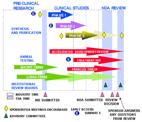
In the post titled: ‘Safety and efficacy of drugs’, we saw how the regulatory agency in the USA, the FDA (Food and Drug Administration), started the drug review process by submiting an Investigational New Drug (IND) application to request the start of clinical trials in humans.

Later, there is a review at two levels:

      A review at national level, where the FDA inspects all the pre-clinic work performed in the laboratory and the data set that led to the submission of that IND.
      A second review at local level (cities, towns, health systems) that is carried out by local entities named Institutional Review Boards (IRBs) regulated by the FDA. Their functions are to evaluate the number of patients that take part in the study, the study safety and its design.
Drug approval process map
After these assessments, if it is concluded that the benefits outweigh the risks the FDA and IRBs approve human trials.

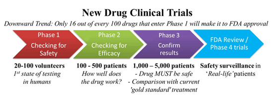
Clinical trials in humans are divided into three different phases:

      Phase I: they are small tests that involve from 10 to 20 healthy human volunteers, who do not have the disease of interest.
In this phase, the drug is, for the first time, administered to human subjects.
The objective of these studies is to find the tolerability and safety of the new product, as well as its pharmacokinetics[1].
      Phase II: once the phase I has been completed without issue, the researchers proceed to the phase II where they determine, by comparison, if the new drug works better than an inactive product (placebo) or, at least, as well as another current drug.
To achieve this, half of the patients of the study are administered with the new drug and the other half with the inactive product or the drug already on the market.
Patients receive a wide range of doses of the drug with the purpose of finding out which one provides fewer side effects along with the best efficacy.
This phase involves between 100 and 500 test subjects.
      Phase III: if everything goes according to plan, we reach phase III where the new drug is given to thousands of patients. Once again, it is compared to a placebo or a medicine that already works to discover whether the new product is safe and efficient, this time at a larger scale.
In this phase, the tests occur in many centres not only in the US but also all over the world.
Another characteristic feature of phase III is that the drug is administered by healthcare providers or practising clinicians, which gives the FDA new data about the safety and effectiveness of the new medicine in the real environment of health care.
New drug clinical trials
In phases I, II, III and even subsequently, the safety of the new compound is always evaluated and is the main concern in these studies.

Biopharmaceutical companies do not conduct these clinical research studies alone, but working very closely with the FDA, in such a way that the FDA can halt a study at any moment, even before beginning, when it suspects that the patient safety could be at risk.
Equally, the FDA can finish a trial ahead of schedule when the drug is so effective that expectations are exceeded, so that the new medicine starts to be commercialised for those people who need it.
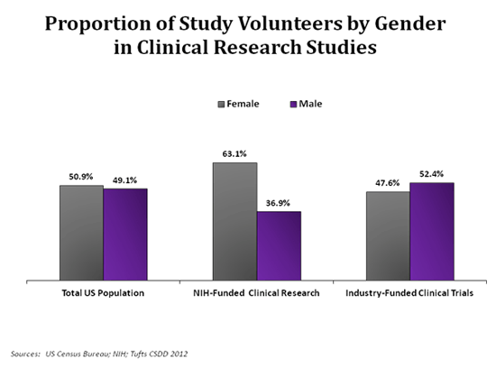
Proportion of study volunteers by gender
At the end of these clinical trials, a new application, named a New Drug Application (NDA), is drawn up and submitted to the FDA where it is examined by several members with different backgrounds: scientists, statisticians, clinicians… in order to give recommendations (as long as the drug fulfils the safety and effectiveness criteria).

The FDA also examines the facilities where the new drug will be manufactured in a standardised and robust way and confirms that they adhere to good manufacturing practices (GMP).
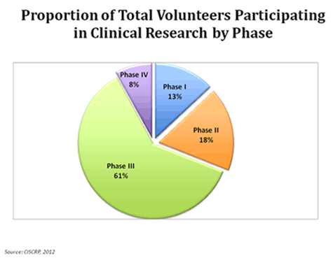
Proportion of volunteers in clinical trials by phase
The FDA also reviews the set of information, called product or package labelling, distributed with the product, which helps patients and health providers to determine if a specific medicine is the most appropriate to treat a certain illness.
This leaflet includes information about the clinical testing done, the most suitable dosage, warnings about its safety (if any) and side effects.

Lastly, the FDA decides whether the new medication is approved or not. If the result is negative, it can ask for further studies from the manufacturer or deny its use in the US permanently in the event that there is enough evidence about the drug’s ineffectiveness or doubts about its safety.


[1] Measurement of drug levels in the organism and how the drug is excreted from it.

Sources: OpenStax College, Biology. OpenStax College. 30 May 2013.
              http://www.ice-epilepsy.org/us-drug-approval-process.html
              http://mscreations.org/clinical-trial-phases/
              https://www.ciscrp.org/education-center/charts-and-statistics/during-participation/


Read more

4.2. Cellular metabolism

0 comments
Practically all tasks performed by living organisms require energy. Energy is not only needed to perform exercise or demanding tasks, but also when we think, and even when we sleep; its use is, therefore, constant.

Energy is used so that immune system cells can absorb, ingest and break down viruses and pathogenic bacteria. It is also fundamental to eliminate toxins and waste, to transport neurotransmitters and hormones and in the synthesis and chemical breakdown of molecules.

Nutrients and other molecules are imported, metabolised (broken down) and synthesised in other molecules, modified in some cases, transported to other cells and probably distributed throughout the body by using energy.

All cellular processes, such as the building and degradation of complex molecules occur through a series of staged chemical reactions.
These chemical reactions within cells, including those that release and use energy, is called cell metabolism.


Metabolic pathways

A metabolic pathway is a series of interconnected chemical reactions that transform one or several substrate molecules gradually, via a group of metabolic intermediates, into a final product or products.

In the case of sugar metabolism, glucose (a simple sugar) is synthesised from smaller molecules:

6CO2 + 6H2 O + energy → C6 H12 O6 + 6O2

While in another different metabolic pathway, glucose is broken down into smaller molecules:

C6H12O6 + 6O2 → 6CO2 + 6H2 O + energy

Hence, metabolism consists of building or anabolism (first reaction) and breakdown or catabolism (second reaction).

These metabolic pathways do not ocurr spontaneously, but each reaction is enabled or catalysed by specific proteins called enzymes, which catalyse both the reactions that release energy and the reactions that require its use.


Potential, free and activation energy

Free and activation energyWhen the bonds in molecules are broken down, they release energy. This is known as potential energy. To carry out their function, cells depend on the extraction of that potential energy.

Free energy is a concept that measures the available energy to perform a task. This free energy is modified during chemical reactions (where energy transfers occur) and this change is denominated as Gibbs free energy (ΔG).
Exergonic and endergonic reactions
The value of ΔG can be negative (whereby energy is released). These reactions are called exergonic or spontaneous. On the contrary, if the value of ΔG is positive then the reaction consumes energy. These reactions are called endergonic or non-spontaneous.

If a reaction is spontaneous, this indicates that its products have less energy than its reactives, and vice versa for non-spontaneous reactions.
Regardless of the sort of reaction, all of them however require an initial contribution of energy to reach the transition state[1]. This contribution is known as activation energy.


ATP: adenosine triphosphate

ATP is the molecule that provides energy to living cells. It is composed of a nucleotide, a pentose (a five carbon atom monosaccharide) and three phosphate groups. The bonds of these three phosphate groups have a high energy content that when are broken, promote a series of reactions and cell processes.
The energy that is released in the ATP hydrolysis reaction:
ATP +H2O → ADP + Pi[2] + energía libre (reacción reversible)
ATP hydrolysis mechanism

Cells use ATP by coupling the exergonic reaction of ATP hydrolysis with endergonic reactions. ATP gives its phosphate group to another molecule during a process known as phosphorylation. This phosphorylated molecule is less stable than its unphosphorylated form, but the added energy after the addition of the phosphate group enables it to perform the necessary endergonic reactions so that the cell can carry out its different functions.


Enzymes

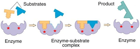
Enzymes (usually one or more polypeptide chain proteins) speed up chemical reactions by diminishing their activation energy requirement.
Enzymes bind to substrates[3] to catalyse reactions in four different ways:
      Taking part directly in chemical reactions by forming transient covalent bonds with substrates.
      Uniting substrates in an optimal orientation.
      Providing optimal conditions so that the reaction can take place.
      Acting on bond structures so that they can be broken more easily.
How an enzyme works

Enzymes are regulated by cell conditions like pH, temperature and their location within the cell (some of them are compartmentalised).
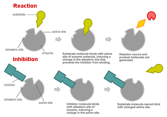
But the most common method by which cells regulate enzymes in metabolic pathways is via feedback inhibition. In this way the products of a metabolic pathway act as inhibitors (normally allosteric[4]) of one or more enzymes involved in the pathway where these products are originated.
Enzyme activity


[1] Very high energy and short duration state that is produced when the reaction reactives approach and experiment a deformation.
[2] Inorganic phosphate group.
[3] Specific chemical reactives on which a specific enzyme performs.
[4] They decrease the enzyme activity by causing structural changes on them, enzyme receptors change and adopt their inactive conformation.

Sources: OpenStax College, Biology. OpenStax College. 30 May 2013.
              http://cienciasdejoseleg.blogspot.com.es/2013/03/plicacion-de-las-variables.html
              http://teenbiotechchallenge.ucdavis.edu/2010_TBC/Peter%20Wang,%20Clara%20Fannjiang,%20William%20Liu/Chemistry.html
              https://mollycools.wordpress.com/2014/03/10/enzymes/
              https://commons.wikimedia.org/wiki/File:Allosteric_competitive_inhibition_3.svg


Read more

4.1.3. Plasma membrane. Cellular transport

0 comments
Among the different functions of the plasma or cell membrane, the most basic is to establish the cell’s limits and keep it functional.
The membrane is selectively permeable. Consequently some elements can enter and exit the cell freely, whereas others need a specialised structure or even energy to accomplish this.

The most significant transport routes that the cell uses through its plasma membrane are:

Passive transport

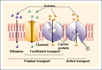
Passive transport is a natural phenomenon where the cell does not need to use any sort of energy to move substances across the plasma membrane.
In passive transport, the substances pass from a high concentration region to a low concentration region.
The various mechanisms of passive transport are:

      Diffusion: a single substance moves from a high concentration area to a lower one until the concentration is equalised on both sides.
A variation of diffusion is the filtration process where materials move through the membrane according to their concentration gradient[1]. At times, the diffusion rate is increased by pressure causing materials to be filtered much more quickly. This is the process that takes place in kidneys where the blood pressure forces big amounts of water and substances dissolved in it (solutes) from the bloodstream to the renal tubules.

      Facilitated transport: diffusion is carried out by the membrane with the help of its embedded proteins, which act like shields for these substances (polar molecules, ions) against the membrane’s repellent forces, facilitating their diffusion inside the cell.

      Osmosis: is a special case of diffusion in which water, not material, is carried across the membrane. Therefore, the membrane limits the diffusion of solutes in water. This process is mediated by a set of specific proteins called aquaporins.
Passive and active transport

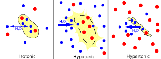
Depending on the relationship between the cell osmolarity[2] and the osmolarity of the surrounding extracellular fluid, we can identify three solution states:

      Hypotonic solutions: the osmolarity of the extracellular fluid is lower than the osmolarity of the fluid inside the cell, therefore water penetrates the cell.

      Hypertonic solutions: the osmolarity of the extracellular fluid is higher than the cytoplasm osmolarity, hence water leaves the cell.

      Isotonic solutions: both the cell and the extracellular fluid have the same osmolarity. As a consequence, there is a constant exchange of water between both regions.
Different types of solutions

Active transport

Active transport, also known as pumps, requires the use of energy in the form of adenosine triphosphate (ATP). This energetic requirement is necessary because the substance is moving against a concentration gradient (i.e., the substance concentration in the cell is higher than in the extracellular fluid or vice versa).
By this mechanism a wide range of different sized molecules can be carried.

In living organisms, there are not only simple concentration gradients but also electrical gradients that indicate a charge difference across the cell membrane.

The cell interior is negative electrically in comparison to the extracellular fluid, which has a higher concentration of K+ (potassium ions) and a lower concentration of Na+ (sodium ions) than the extracellular fluid.
Consequently, the electrical and concentration gradients of Na+ tend to lead these ions into the cell.
The K+ ion situation is more complex; while its electrical gradient also tends to drive these ions towards the interior of the cell, its concentration gradient leads out of the cell.

These combined gradients of electrical charge and concentration that affect an ion are named electrochemical gradient.

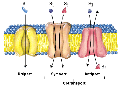
A notable adaptation of the cell membrane for active transport is the presence of specific transport proteins (pumps) that enable the movement of materials on both sides of the membrane. These proteins or transporters are classified into three types:

- Uniporters: they carry a specific molecule or ion in just one direction.
- Symporters: they transport two different ions or molecules in the same direction.
- Antiporters: they also transport two different molecules or ions but in opposite directions.
Classification of transporters

Primary active transport
This type of transport works with the active transport of K and Na and enables secondary active transport to occur at later stage.

One of the most important mechanisms of active transport in animal cells is the Na+ -K+ pump that maintains the electrochemical gradient and the appropiate concentrations of Na+ and K+ in cells.
During this process for every three Na+ ions that are expelled from the cell, two K+ ions penetrate into the cell, therefore the interior of the cell is slightly more negative than its exterior. This charge difference is vital to create the necessary conditions for the second process.

The Na-K pump belongs to the electrogenic pump category, which is a kind of ionic pump that generates a potential electrical charge difference on both sides of the cell membrane.

Secondary active transport (co-transport)
Via this process Na+ ions and other compounds penetrate into the cell taking advantage of the electrochemical gradient created in the previous step.
In this class of active transport a solute that moves against the concentration gradient is co-transported with other solutes that move in favour of its concentration gradient.
Thanks to this method, many diverse molecules, like amino acids or glucose molecules, can enter the cell.
This process is also used to store hydrogen ions of high energy in mitochondria to produce ATP.


Bulk transport

In addition to small ions and molecules, cells must also assimilate and eliminate larger size molecules and particles and in order to achieve it they need a supply of energy. However, these molecules are so large that not even with the supply of energy can they pass through the plasma membrane.
Hence, the cell uses the following mechanisms:

Endocytosis
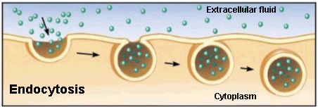
Endocytosis process
Endocytosis is a type of active transport where particles, such as large molecules, parts of cells and even entire cells are enclosed by invagination of the membrane forming a vesicle whose content is carried from the outside to the inside of the cytoplasm.

Listed are the various endocytosis:

      Phagocytosis: is the process in which cells, or large parts are ‘consumed’ by a cell. This is the method used by a particular type of white cells called neutrophils. These immune system cells surround, cover and destroy the microrganisms that invade our body.

      Pinocytosis: the cell captures the molecules that are needed from the extracellular fluid. The cell membrane invaginates that unwelcome cell, creating a vesicle around the liquid of the external fluid, which is ‘consumed’ and released in the cytoplasm.
For example, the maturation egg process in the uterus is carried out by pynocytosis when the ovule takes in the nutrients that its supply cells release.

      Receptor-mediated endocytosis: uses receptor proteins from the cell membrane that have a specific affinity to binding to particular substances.
Endocytosis in animal cells

Some human diseases are caused by the malfunction of this mechanism. It is the case of familial hypercholesterolemia, in which the receptors of bad cholesterol are faulty or completely absent.

Exocytosis
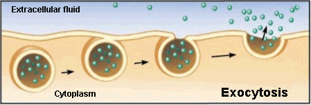
Exocytosis process
Exocytosis is the opposite to endocytosis in which the cell expels material from its interior to outside.
This waste material is covered with a membranous sac that fuses to the plasma membrane and then opens to release its content into the extracellular space.
The neurotransmitter secretion by vesicles in the synaptic gap of neurons follows this process.


[1] Different concentration of molecules between two regions.
[2] It describes the overall concentration of solute in a solution.

Sources: OpenStax College, Biology. OpenStax College. 30 May 2013.
              http://www.educ.ar/sitios/educar/recursos/ver?id=14378
              http://sevendaysperweek.blogspot.com.es/2015/09/spm-biology-3-movement-of-substances_20.html
              http://cbc.arizona.edu/classes/bioc462/462a/NOTES/LIPIDS/transport.html
              Nelson & Cox, Lehninger Principles of Biochemistry, 3rd ed., 2000
              http://es.slideshare.net/exitoinevitable/clulas-31270712
              https://micro.magnet.fsu.edu/cells/endosomes/endosomes.html


Read more

1.4.2. Good Clinical Practice (GCP)

0 comments
According to the European regulations, all clinical trials must be performed in accordance with the rules of good clinical practice (GCP) that constitute a guarantee for patient protection and for test results.

That GCP guidelines of the International Conference on Harmonisation (ICH)[1] have been followed since 1996 in Europe; they have also been adopted by the USA and Japan. They are a set of scientific and ethical quality requirements recognised internationally that must be fulfilled by the planning, execution, record and communication of clinical trials where human beings take part.
These rules must guarantee the rights, safety and well-being of trial subjects and the reliability of the results.

GCP arises from the need to reconciling the subject’s interests (who receives the treatment) and society’s interests in increasing its knowledge about drugs.

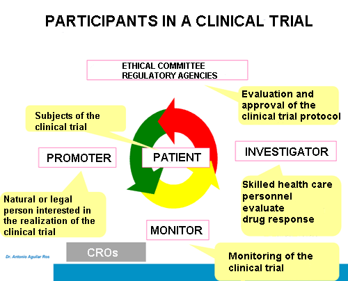
The main actors that participate in a clinical trial are the following:

      Promoter: legal entity or person interested in performing a clinical test and responsible for the same. It is usually a pharmaceutical multinational.
      Ethical committees: they belong to the centre where the research takes place.
      Regulatory agencies: they are usually QUANGOs with government oversight.
These two last participants must approve the protocol and supervise the experimental development. In addition, agencies evaluate the research results to authorise subsequent drug commercialisation.
      Researchers: carry out or manage the test research. They are normally physicians or health professionals that examine the compound response that is subject of study.
      Monitor: responsible for the direct follow-up of the clinical trial execution. Monitor is the intermediary between promoter and researcher and must guarantee the tracking of everything that occurs throughout the experiment.
      Patient: subjects who take part in the research and generally suffer from the disease of interest.
On occasion, some companies called CROs (contract research organisations) provide the trial management and monitoring.
Participants in a clinical trial

After the II World War, the Nuremberg Code was promulgated to establish clinical research principles in human beings, which are as follows:

      Autonomy: respect the principle for subjects that they are treated as autonomous people to decide freely if they want to take part in the clinical test.
The informed consent[2] signed by the patient is part of the fulfilment of this principle.
      Well-being: the physician is obligated to do good and to attend to the welfare of the patients, as well as their highest benefit.
      Non-malefience: to avoid harmful clinical tests for patients.
      Justice: obtained positive results must be shared with the people who suffer from the malady which is the focus of the trial (whether knowledge or new medication). This principle would not be fulfilled, for example, if a country from the third world, where the trial takes place, would not benefit from the treatment because of its high cost.


Phase IV clinical trials

Once a drug has been introduced to the market, studies continue to increase the knowledge about its safety and effectiveness, identify new risks and non-detected adverse response in previous research.
The group of controlled and randomised trials and studies that are performed at this given point is called phase IV of clinical trials.
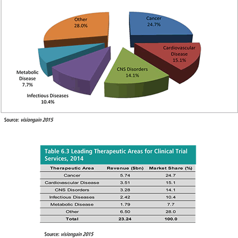
Leading therapeutic areas for clinical trials services

The most common epidemiological (observational) studies after the drug authorisation are case-control and cohort studies.

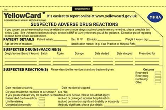
Finally, the Yellow Card Scheme is the system for collecting suspected adverse reactions to drugs. This system is used especially when a medicine has just been marketed and it is mandatory for physicians (and current patients can also fill it out).
Yellow Card for suspected adverse reactions to drugs in the UK

These cards consist of several sections among which we find the following:

      Patient details: first name or initials, age, sex, weight, height…
      Data about the suspected medicine causing adverse reactions.
      Description of those reactions.
      Reporter/clinician details: name, professional address, specialty…


[1] It matches the different regional requirements for the register of pharmaceutical products.
[2] The subject who participates in the clinical trial is informed by this document about the details, advantages and disadvantages of the test with an accessible language, being able to leave it freely at any time.

Sources: CEU Universidad San Pablo: Farmacología Básica, 2013.
              https://www.gov.uk/guidance/the-yellow-card-scheme-guidance-for-healthcare-professionals
              https://yellowcard.mhra.gov.uk/the-yellow-card-scheme/
              http://www.ich.org/home.html


Read more