• Biology

    Biology

    The atom, biological macromolecules (carbohydrates, lípids, proteins, nucleic acids), cell biology, cancer

  • Drugs

    Drugs

    Drug development, pharmacodynamics, pharmacokinetics, toxicology

  • Nanomedicine

    Nanomedicine

    Introduction to nanotechnology, diagnostic devices, drug delivery, regenerative medicine

  • Miscellaneous

    Miscellaneous

    A more in-depth explanation of topics previously mentioned in the other sections

Posts for label: Nanomedicine

Posts for label: Nanomedicine

2.2.3. Nanobiosensors

0 comments
The great potential that nanotechnology offers enables the progress of new devices in the nanodiagnostic field, mainly in nanobiosensors.
Their properties are characterised by the nanoscale at which they are produced. Unlike biosensors , which we studied in the post: "Introduction to biosensors", where their sensory receptors are distanced by hundreds of micrometers, here they are distanced by a few nanometers.
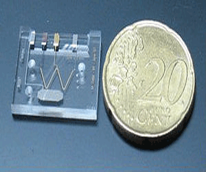
Actual size of a lab-on-a-chip
Thus, even sensors constituted by single molecules bound to their surface have been designed. Consequently the process of diagnosis is performed with a much smaller size device, improving portability and the potential for use anywhere.
Also, only extremely small sample quantities (micro or nanolitres) are required to carry out the analysis, so the sample extraction methods are less traumatic and invasive for patients.

These instruments also facilitate the use of samples without fluorescent or radioactive markers, which are used in biological and clinical analysis. This property is essential, since it has been proven that when a sample is not marked or altered before examining, the detection sensitivity significantly increases.

Within nanobiosensors we can distinguish the following categories:


Nanophotonic biosensors

This class of nanobiosensors, also known as evanescent wave biosensors, is based on how the light is transmitted by multiple internal reflections through the optical waveguide, which is a component of the biosensor.
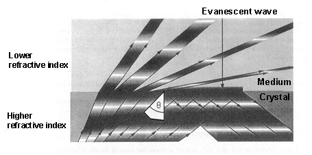
Evanescent wave productionWhen light is propagated in a medium and passes into a lower refractive index[1] medium, it is not totally reflected. As a consequence, a light component (called evanescent wave), in each of these reflections, is propagated in the lower refractive index that covers the waveguide. This propagation is really short, a few hundreds of nanometers, but it permits interaction between the light and the detection surface of the sensor, demonstrating which specific bioreceptors anchored to that surface interact with the analysed sample.

With these devices, only a few microliters of sample are necessary to determine protein concentrations or the variations of just one DNA base.

Also, it is possible to evaluate the metabolic state of just a single cell because some of these nanobiosensors have sharp fibre optic (30-50 nm) filaments, which can be introduced into cells through the plasma membrane without harming the cell.
This method facilitates the study of in vivo cell functions (apoptosis[2], cell division, biological nanomachines…) and it can detect pathological changes in a single cell.


Nanoplasmonic biosensors

In the Surface Plasmon Resonance (SPR) biosensor, a thin metal layer (normally a 50- nm-thick layer of gold) is placed on a dielectric material (a crystal), and its functioning is based on the detection of changes in the refractive index around the separation area between both elements.
By exciting the interface between these two layers (under conditions of total internal reflection) a plasmonic resonance[3] is activated with a certain angle of incidence.. This angle, which produces a plasmonic wave (with an evanescent character) is very sensitive to the changes from molecular interactions that happen on the metal surface. Therefore, these interactions between the analyte[4] of the sample and the sensor surface of the biosensor are shown as variations of the resonance angle.
Generation of Surface Plasmon Resonance (SPR)

Currently, new biosensors are being developed, which rely on the plasmon resonance phenomenon in nanoparticles. While their detection range would be very similar to the SPRs, their functioning system can be made simpler by measuring light transmission instead of light reflection, in addition to the advantage of the device miniaturisation.

Due to the tiny size of nanoparticles, the electron oscillations are more localised than in the previous case (SPR), in specific areas of nanoparticles. This phenomenon is called ‘Localized Surface Plasmon Resonance’ (LSPR). The colour (wavelength: λ) changes that nanoparticles experience by adsorbing[5] the (bio)molecules of the examined sample are used to analyse this sample.
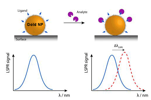
Variation of wavelength (Δλ) in nanoparticles when LSPR is produced

An alternative, within this kind of device,, is DNA sensors detecting colour changes produced by the aggregation of gold nanoparticles marked with stranded DNA complementary to the target DNA.

Nanoplasmonic biosensors are characterised by their real-time detection, specificity, high sensitivity and recognisition rate.
Their main applications are found in veterinarian, biomedical and environmental fields as well as in the food industry.


Nanomechanical biosensors

These sorts of nanobiosensors use the deflection or the resonance frequency variation of a microcantilever (as a transducing[6] method) when they interact with the studied sample. This position variation (Δx) is just a few nanometers derived from the biomolecular identification of the analysed sample.
Working principle of nanomechanical biosensors

Because these microcantilevers can be produced in mass at a low cost by standar microelectronic technology, thousands of them can be manufactured to identify thousands of analytes in a single sample.

The sensor area of these microcantilevers is around 1,000 μm2, which provides them to test sample volume lower than femtomole (10-15 moles).

Both photonic and nanomechanical biosensors could help us to obtain unlimited genetic and proteomic information that would offer the discovery of pathogens, new drugs, vaccines and undiscovered mutations of certain diseases in a faster way than the current technology.


Microfluidic devices or lab-on-a-chip (LOC)

This class of instruments uses an electric field to move liquids, particles, molecules or cells through microcapillaries designed on chips of different materials like silicon, crystal, quartz or plastic.
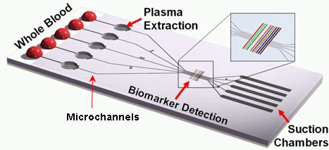
A schematic diagram of a  lab-on-a-chipThis kind of chip is configured by a large number of integrated microchannels and microchambers, where the examined sample is subjected to complex chemical and biochemical reactions.
The required volume of the sample fluids is very small, therefore the analysis can be carried out very quickly.
It is a portable and disposable detection method with a high grade of automation.

The ultimate objective for all these biocompatible nanodevices is to finally be implanted in vivo inside our organism, where they would perform a similar function to ‘sentinels’ in the presence of the first diseased cells. Some advances have already been made to that effect at the microscale (pills with built-in image cameras), but, without a doubt, this will become one the major research fields in nanomedicine in the near future.


[1] It describes how light propagates through a certain medium. It is defined as n = c/v, where c is the speed of light in vaccum and v the speed of light in that medium.
[2] Process of programmed cell death.
[3] Excitation or oscillation of conduction electrons at the interface between two materials, in our case a metal layer on a dielectric substrate.
[4] Substance (ion, compound or element) of interest in an analysed sample.
[5] Adhesion of atoms, ions or molecules to a surface.
[6] Process by one signal or energy is converted to another.

Sources: Nanomedicina: aplicación de la nanotecnología en la salud. Laura M. Lechuga. Grupo de Nanobiosensores y Aplicaciones Bioanalíticas
              Centro de Investigación en Nanociencia y Nanotecnología (CIN2). CSIC
              http://www.kennislink.nl/publicaties/de-opmars-van-de-twentse-lab-on-a-chip
              http://images.slideplayer.es/16/5040279/slides/slide_47.jpg
              http://www.mdpi.com/sensors/sensors-10-09630/article_deploy/html/images/sensors-10-09630f1-1024.png  
              https://www.ifm.liu.se/applphys/molphys/research/biosensing_using_nanopart/
              http://www.tcd.ie/Physics/people/Martin.Hegner/ReviewNSST-The_impact_of_STM_and_AFM.html?ntherodt_01.pdf
              Fritz, J., Baller, M.K., Lang, H.P., Rothuizen, H., Vettiger, P., Meyer, E., Guntherodt, H.-J., Gerber, CH. and Gimzewski, J.K., 
              Science 288 (2000) 316.
              https://www.theengineer.co.uk/lab-on-chip-device-promises-hiv-diagnosis-in-10-minutes/


Read more

2.2.2. Imaging diagnostics

0 comments
Magnetic resonance scannerOver the last few years, imaging diagnostics has significantly increased its relevance such that they have become an indispensable tool to diagnose innumerable diseases such as neurological syndromes, cardiovascular disorders or cancer.

The convergence between imaging diagnostics and nanotechnology enables the creation of a new series of tracers and contrast agents with a higher resolution and sensitivity, which allows illness detection at earlier stages at cellular or even molecular level. This advance makes the quick application of treatment possible, thus increasing the chances of treatment and survival.

Imaging diagnostic nanosystems are mainly based on three types of nanoparticles: semiconductor, metal and magnetic.


Semiconductor nanoparticles

This first sort of nanoparticles, also called quantum dots, consist of semiconductors[1], whose size has been reduced to a few nanometres (1-10 nm). This produces changes in their electronic arrangement, which means they lose their distinctive band structure.
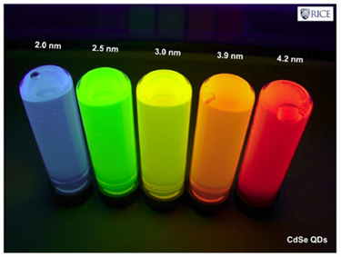
Because of this new structure, their optical response (specifically their fluorescence) is going to be closely related to the size variation of these nanoparticles. Consequently, these quantum dots emit light in a broad range of wavelengths (colours), depending on their size and not the material they are made of, which turns them into excellent biological markers.
Relationship between quantum dot size and wavelength emitted
While a wide variety of semiconductor materials have been examined, the most commonly used are cadmium selenide (CdSe) and cadmium telluride (CdTe), because in addition to be manufactured on a large scale, their sizes can be controlled perfectly. This enables us to see emission bandwiths with a full spectrum of different colours over a long lifetime.

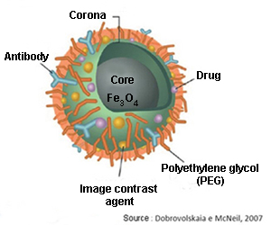
But the semiconductor nanoparticles must also get to their defined targets. To achieve that, the quantum dots must be coated with polymers, like polyethylene glycol (PEG), which makes them invisible to macrophages and prevents immune system cells degrading and digesting them before reaching their target.

The next step is to make these nanoparticles able to identify their targets through bioreceptors (antibodies) that are bound to their surface, and these in turn will be bound to the specific antigens[2] of the target cells (e.g.: cancer cells). The bioreceptors of quantum dots produce a biomolecular recognition reaction when they meet these antigens, being accumulated in that area, and they are observed by using ultraviolet light due to the peculiar fluorescence that they emit.

Lastly and once they have fulfilled their role, the quantum dots must be purged from the organism to avoid unwanted side effects. It seems that the quantum dots are excreted through the kidneys and the liver without difficulties in animal testing, but to be tested in humans some problems related to the aggregation process must be resolved before receiving authorisation from the health agencies for commercialisation.


Metal nanoparticles

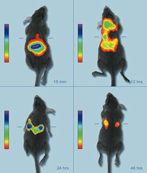
A live mouse model showing the accumulation of near infrared fluorescent nanoparticles in breast cancer tumorsThe fact that metal nanoparticles have a resonance frequency (their colour) dependent on their size and shape makes them a second option for contrast agents.
This property provides enables them to be manufactured to absorb or reflect light in the spectrum of interest. Thus, gold nanoparticles can be designed to absorb or reflect light in the near-infrared band (700-900 nm) as biological tissues are more transparent in that electromagnetic spectrum band. Using techniques like optical coherence tomography (OCT)[3], 3D maps are obtained of the areas where the nanoparticles are gathered.


Magnetic nanoparticles

A third alternative is magnetic nanoparticles (iron oxides like magnetite: Fe3O4), with the ability to increase the contrast in magnetic resonance imaging (MRI) tests.
Functionalized magnetic nanoparticlesTheir transport through the organism can be carried out with the use of an external magnetic field (like an electromagnet) taking advantage of their magnetic properties. They may replace the old fashioned markers made of heavy metals in the near future, because they have lower toxicity.

Both magnetic and metal nanoparticles use identical or very similar methods to the ones described for the quantum dots in order to avoid degradation by the immune system, thus enabling them to locate and bind to the target tissues and cells.

Imaging nanosystems belong to the in vivo diagnostic category, therefore they must be injected into the human body. This implies, as we have seen, potential drawbacks (biocompatibility, sophisticated design) that have to be resolved to ensure an effective and safe use in the organism, minimising undesirable side effects.


[1] Material that behaves as an insulator or a conductive material depending on several factors such as environment temperature, pressure, surrounding electrical and magnetic fields or the incident radiation.
[2] External or toxic substances for the body (generally proteins) that lead to antibody production and causing an immune response. 
[3] Non-invasive imaging test that uses light waves to take cross section images.

Sources: Informe de vigilancia tecnológica: nanomedicina. Fundación para el conocimiento madri+d. CEIM. José Manuel González, Marta López, 
              Gema Ruiz.
              Nanomedicina: aplicación de la nanotecnología en la salud. Laura M. Lechuga. Grupo de Nanobiosensores y Aplicaciones Bioanalíticas
              Centro de Investigación en Nanociencia y Nanotecnología (CIN2). CSIC
              http://www1.radiology.ucsf.edu/research/labs/hyperpolarized-mri-tech-2/facilities_equipment
              http://www.osiconference.org/osi2015/presentations/Tu2.3%20Zahn.pdf
              http://www.nanowerk.com/news/newsid=7441.php (Image: Penn State)
              http://slideplayer.com.br/slide/74350/


Read more

2.2.1. Introduction to biosensors

0 comments
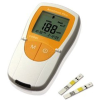
As a consequence of our current technology, the diagnosis of certain diseases is made at overly advanced stage of the disease.Example of biosensor: glucometer
Today, the main instrument used to diagnose illnesses is the biosensor, which enables the detection of specific substances, their composition and molecular features, or to identify the presence of microorganisms in certain environments.

This instrument is comprised of two main components:

       A reactive or sensory area formed by a biological receptor (antibodies, DNA, enzimes…) with the ability to join those substances to be detected.
      A transducer system that processes and quantifies the signal (mechanical, electrical, optical…), which is caused by the interaction between the analysed substance and the recognition element.
The integration of both components makes biosensors sensitive and selective.
General scheme for biosensors
Biosensors have a wide range of applications in fields like environment, healthcare, biotechnology and the food industry.

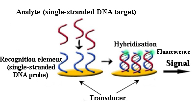
Among the most common biosensors we find microarrays or biochips which consist of hundreds or thousands of biomolecules (generally oligonucleotides[1] or single-stranded DNA fragments, often known as ‘bioreceptor probes’) located on a solid surface (glass, silicon, gold) in particular positions and concentrations making a 2D matrix, with each molecule separated by a distance of between 100 and 150 μm from each other.
Biosensor for DNA detection
The examined sample is marked (normally with reactive fluorescent dyes), in such a way that, when it comes into direct contact with the sensory molecules of the biosensor, they are hybridised[2] with their homologous sequences, producing a fluorescent signal in that location.
Later, the data obtained are analysed and interpreted using a scanner and computer tools.

Biochips have countless applications: from personalised medicine (to know our higher or lower predisposition to suffer cancer or specific genetic disorders), the detection of harmful bacteria in food or water, discovery of virus and bacteria mutations, which become drug resistant or examining the microbial diversity in certain environments.

Another type of microarray are the protein microarrays (or protein chips) which, in contrast to DNA fragments, have thousands of functionally active proteins anchored to their surface. This class of instrument has great potential in basic research applied to molecular biology, the identification of disease markers and the search for therapeutic targets.

Lastly, we find within microarray technology cell chips. Antigens, proteins or even lipids are placed on these detector surfaces, which interact with cells, not only capturing them, but also triggering responses on them, such as phenotypic changes[3] or the segregation of particular substances.
These kinds of microarrays are used for toxicological analysis, identification of illness markers and the analysis of pathogenic agents.
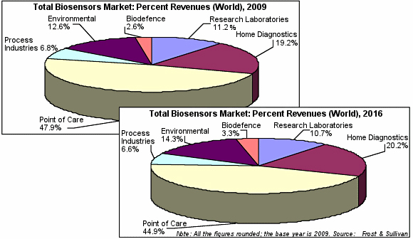
Evolution of total biosensors market: 2009-2016

Science keeps developing and it is designing smaller and more powerful devices, on the basis of the enormous potential that nanotechnology offers. These diagnostic nanosystems, whose main advantage is, in comparison to current biosensors, the early detection of diseases, depending on their working area, are divided into: imaging diagnostic nanosystems and nanobiosensors, which will be object of study in the coming posts within this section.


[1] General term for a short, single stranded DNA or RNA used in research, genetic testing and forensics.
[2] Recognition and combination of two complementary molecules
[3] Changes in those particular and genetically inherited features of an organism. 

Sources: Informe de vigilancia tecnológica: nanomedicina. Fundación para el conocimiento madri+d. CEIM
              Fundación española para la ciencia y la tecnología (FECYT). Nanociencia y nanotecnología. Entre la ciencia ficción del presente 
              y la tecnología del futuro, 2009.
              http://www.definicionabc.com/ciencia/fenotipo.php
              http://www.blogdefarmacia.com/biosensores-tecnologia-para-la-salud/
              http://www.pharmatutor.org/articles/applications-of-biosensors-technology-future-trends-development-and-new-intervation-in-                                   biotechnology?page=0,1
              http://www.slideshare.net/ManjuAnshika/biosensor-dr-manju-jha
              http://www.sensorsmag.com/specialty-markets/medical/strong-growth-predicted-biosensors-market-7640


Read more

2.1. Introduction to nanomedicine

0 comments
The application of nanotechnology in the healthcare field has enabled the development of a new scientific discipline known as nanomedicine.

Due to the fact that some of the fundamental biological macromolecules of living organisms (like the DNA and proteins) are located at nanoscale, this has allowed the interaction between nanometric devices and nanomaterials within the human body. This has facilitated the discovery of important new advances in medicine.

Certain predictions made in the early stages of the development of nanotechnology are still science fiction.
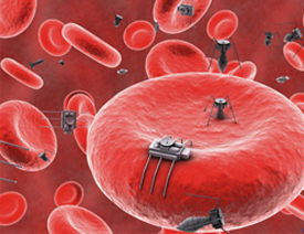
Imaginary nanobots in the bloodstream
For instance, the idea that we could develop “nanobots” that would protect us against external microorganisms, would cure injuries and damaged tissues selectively. Nevertheless, some major advances have been made.

The steady increase of neurodegenerative and cardiovascular diseases, diabetes or cancer  requires us to investigate new diagnostic and therapeutic techniques that must be simpler, quicker and more precise than the ones we have today, decreasing, at the same time, the costs involved. It is expected that nanomedicine should address some of these problems, like diagnosing disease at its earliest stage, tailored treatments for patients or the ability to regenerate damaged organs and tissues.

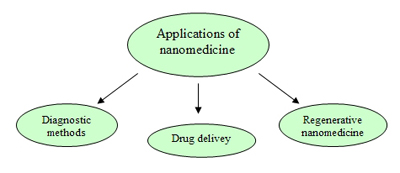
Nanomedicine is focused on three large areas: diagnostic methods, drug delivery systems and regenerative medicine. Nanodiagnostic techniques are divided into analysis systems and methods of medical imaging. Both methods enable the detection of the appearance of the disease, in vitro and in vivo at early stages.  This makes it possible to take action with the most appropiate treatment and causing as little damage as possible to the rest of the body.
Fields of applications of nanomedicine
In sample analysis of patients, nanomedicine also helps to achieve a more specific, quicker and more effective diagnosis. In this way diseases are diagnosed at cellular and molecular level providing more chance of a cure.
Nanomedicine has also advanced drug delivery systems: drugs are guided specifically to those affected areas and cells to accomplish a more effective treatment with a lower dose and minimising side effects. In addition, these systems protect the carried drug by avoiding degradation of the drug before it reaches its target. Thus, the challenges of  delivery associated with drugs with poor solubility or with those that cannot be delivered using conventional methods is solved.
Lastly, through gene and cell therapies, biomaterials, tissue engineering and nanotech tools, regenerative nanomedicine boosts the natural repairing mechanisms of the human body which often are not enough to achieve the total recovery of the organism alone.


Sources: Informe de vigilancia tecnológica: nanomedicina. Fundación para el conocimiento madri+d CEIM
              Nanomedicina: aplicación de la nanotecnología en la salud. Laura M. Lechuga.Grupo de Nanobiosensores y Aplicaciones Bioanalíticas
              Centro de Investigación en Nanociencia y Nanotecnología (CIN2). CSIC
              Fundación española para la ciencia y la tecnología (FECYT). Nanociencia y nanotecnología. Entre la ciencia ficción del presente y la                     tecnología del futuro, 2009.


Read more

1.3. Top-down and bottom-up approach

0 comments
Current manufacturing technology is based on the approach known as “top-down” which consists of, in a similar way to a sculptor does, starting from a large block and shaping and chiseling it, to obtain, progressively, a smaller object with a desired shape.
In the nano approach, we progress in the opposite way, from the small structure, building up to a bigger one. This method is called the “bottom-up” approach, in which like lego (with a large number of pieces with different shapes, colours and size), we are going to begin with basic elements such as atoms, nanoparticles, nucleic acids or proteins to assemble molecules or even to build diverse sensors and devices.
To enable manufacturing using nanotechnology, special tools which enabled the visualization as well as the manipulation of objects at nanoscale are required. The scanning probe microscopy (SPM), for instance, accomplishes this task by allowing us not only to observe but also to move atoms on a surface.

The progress of nanoscience and nanotechnology and the use of this highly advanced instrumentation created one of the most significant features of this field: it is multidisciplinary. By reducing the scale, atoms and molecules become the basic “bricks” which physicist, chemists, biologists and engineers work with, using a common language. One example of this multidisciplinarity is the design and manufacturing of a biosensor where a biologist must have knowledge of quantum physics and a physicist about biology in order to design their end product successfully.

Assembly on a molecular or particle basis to develop all the technology we demand may seem unrealistic, nevertheless this is what the Earth has been doing for the last 4 billion years, since starting from simple molecules it formed really complex structures by linking and auto-assembling substances. Any living organism is, undoubtedly, a clear example of bottom-up building; starting from certain organic molecules and a genetic sequence it has been possible to create very complex structures functionally and structurally. As a consequence, nanotechnology can learn from these processes to imitate and adapt them, even, to other kinds of problems very different to biology.

The development of nanoscience and nanotechnology, according to experts, will occur in three stages. The firt one between the years 2000 and 2020, where industries will mainly keep using conventional production techniques (top-down). The second stage between 2010 and 2030, where bottom-up methodology will begin to spread, and ultimately become the leading manufacturing scheme for the rest of the XXI century. But this fact does not mean that our current procedures will totally disappear, because the use of one system or the other will be dependent on many factors like raw materials, labour, environmental and social costs and, of course, economic profitability.
Nanotechnology is already a major industry, worth 50 billion dollars globally market in 2006, with expected growth to a trillion dollars in 2015. Therefore, this market will benefit those companies whose countries are investing in this field long term.


Sources: Fundación española para la ciencia y la tecnología (FECYT). Nanociencia y nanotecnología. Entre la ciencia ficción del presente 
              y la tecnología del futuro, 2009.
              http://researcher.watson.ibm.com/researcher/view_group_subpage.php?id=4252
              http://www3.nd.edu/~kamatlab/facilities_physchar.html


Read more

1.2. Nanostructures

0 comments
Nanostructures can be defined as those objects where, at least one of their dimensions is located at nanoscale (1-100 nm). Among the most commonly used structures in nanomedicine field we find:

      Micelles: structures with spherical or globular shape. They are made of molecules that have a polar or hydrophilic head (with strong affinity for water) and a non-polar or hydrophobic tail (repels water). The heads are placed in the outer region of the micelle forming a layer in contact with the liquid environment surrounding it; the tail, instead, is located in the interior forming the nucleus of this nanostructure.
Their typical size is c50 nm and they are used to carry and deliver water-insoluble drugs, which are confined inside the hydrophobic core of the micelle, in such a way that they are protected from the exterior aqueous environment.
One of their most interesting features is that they can circulate through the bloodstream for longer than other sorts of particles because they can avoid macrophage[1] action.

      Liposomes: closed vesicles made up of lipid bilayers (two lipid layers faced by their hydrophobic tails). According to the number of these bilayers the liposomes may be classified as unilamellar or multilamellar.
Unilamellar liposomes are characterised by an aqueous core to carry water soluble drugs. Multilamellar liposomes do the same thing with liposoluble drugs. When they are intravenously administrated, liposomes are cleared quickly by the reticulendothelial system (RES)[2], in addition, hydrophobic, electrostatic  and Van der Waals forces may disintegrate them. To avoid this, these nanostructures are coated with inert polymers, like polyethylene glycol (PEG) which enables their circulation through the body without being excreted.
Some liposomes are designed to be degraded only where necessary, for instance, in low pH areas (tumour regions with hypoxia[3]), others are functionalized with antibodies or ligands (molecules that bind specific celullar receptor) onto the nanoparticle surface so that they can bind and perform on these receptors. A third technique is based on the development of thermo-labile liposomes, which are guided to target tumor tissue by hyperthermia.

      Dendrimers: three-dimensional systems with treelike structures. These nanostructures consist of a central core molecule with lots of branches. Their shapes and sizes are controlled in a very accurate way by polimerization from the central core or by synthesis from the periphery to the core molecule.
Dendrimers are excellent candidates for carrying drugs, due to the fact that they offer a high stability and their functionalization (attachment of functional groups on the surface of the nanoparticle) by physical or chemical interactions.
They can deliver a wide range of molecules, both hydrophilic and hydrophobic ones, anticancer agents, drugs or contrast agents for diagnostic imaging.
Types of nanostructures

      Nanospheres: spherical structures made of matrix systems in which the drug is distributed by encapsulation, entrapment or attachment. The nanosphere surface is modified by the addition of biological material (antibodies or ligands) or polymers so that they can reach their targets in cells.

      Nanocapsules: vesicle systems where the drug is confined in a cavity or nuclear core, surrounded by a polymeric membrane where ligands and antibodies can be attached. The core material may be solid, liquid or even gas, always in an aqueous or oily environment.

      Carbon nanotubes: mono or multilayer cylindrical structures constituted by graphite or another carbon material. They deliver their loads in a specific way by functionalizing their surfaces with nucleic acids, proteins or bioactive peptides[4], which allows them to become particles with a very low toxicity. They are good candidates for carrying and delivering drugs because they are not immunogenic (they do not produce an immune response).

      Polimeric nanoparticles: are one of the most adequate and suitable material used in nanomedicine, being largely biocompatible and biodegradable. Among their advantages are: the potential to modify their surfaces by chemical transformations, the encapsulation of the load to transport, carrying a broad variety of therapeutic agents and lastly an outstanding pharmacokinetic control[5] of these agents.
Their polymeric coating reduces immnunogenicity and limits their phagocytosis by the reticuloendothelial system (RES), increasing, in this way, the blood levels of the carried drugs in organs like the brain, intestines and kidneys. They are normally designed in such a way that are sensitive to the environment, delivering their carried drugs by responding to both physical stimuli (temperature, solvents, light) and chemical stimuli (reactants, pH, ions in solution or chemical recognition).

      Inorganic nanoparticles: usually composed by (SiO2) or alumina (Al2O3).  However, their cores are not just limited to these two materials, they can be made of any kind of metal, oxides and metal sulfides, which leads to a myriad of nanoparticles with a large range of shapes, sizes and porosities.
They are normally produced to avoid RES by modifying their size and superficial composition. They are porous with a physical coating that protects the carried load from a likely degradation or denaturation.


[1] Large size cells of the immune system located in different organs.
[2] System composed by a group of cells whose function is to capture inert particles in the body.
[3] Condition in which blood, cells or tissues is deprived of sufficient oxygen supply.
[4] Molecules formed by the union of several amino acids.
[5] Control of the processes that a drug is undergo within the organism.

Sources: Barbara Haley, Eugene Frenkel, Nanoparticles for drug delivery. Elsevier, 2008.
             Jose Manuel González, Marta López, Gema Ruiz. Informe de vigilancia tecnológica, nanomedicna 2006
             Amir H. Faraji, Peter Wipf. Nanoparticles in cellular drug delivery. Elsevier, 2009


Read more

1.1. Nanotechnology. Introduction

0 comments
It is very likely that the term nanomedicine has caught your eye and maybe this is the first time you have heard of it. As nanomedicine can be defined as the application of nanotechnology in the medical field, I will begin this section with a brief view about what nanotechnology is. I will show you the unique features of technologies and materials at nanoscale and a classification of the most common types of structures used in nanomedicine.

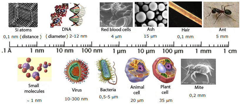
But first of all, what is a nanometre?, the prefix “nano” comes from the Greek νάνος, which means “dwarf”. Nowadays, it indicates the billionth part or, in other words, it is a factor of 10-9, therefore, a nanometre (nm) is one billionth of a meter. This way, we can conclude that nanoscience/nanotechnology is the science/engineering that studies and operates with matter in a size from 1 to 100 nm.

The idea and concept of nanotechnology arose from a Richard Feynman’s conference at UCLA University in 1967, where this theoretical physicist introduced, for the first time, the possibility of manipulating atoms and molecules.
But nanotechnology age really began in 1981 when the scanning tunneling microscope (STM) was developed with which we are capable of observing atoms.
The smallest objects we can observe with our naked eye have a size around a millimetre (the thousandth part of a meter), e.g.: the edge of a coin or a grain of sand, below this magnitude we find it difficult to distinguish objects.
If we divide a millimetre into a thousand parts we are in the micrometer scale, which is the domain of bacteria (5-20 μm) or blood cells (red blood cells: 6-10 μm). Therefore, to observe them we need the help of an optic microscope.
If we keep decreasing the scale and we cut up a micrometer into a thousand parts, we achieve our goal: the nanometre, as I said previously the billionth part of a meter. At this scale we find viruses (20-250 nm) and the DNA molecule (around 2 nm wide).
However, to be able to observe an atom we should still decrease one order of magnitude our scale, as atoms have magnitudes from 0.1 to 0.3 nm.
The scale of things
To get a more accurate idea about the real size of a nanometre, here are a couple of objects from our daily life measured in nanometres: a human hair is about 50,000-100,000 nm in diameter and a paper sheet is around 100,000 nm thick. Now, let’s do it the other way round, imagine you are shrunk until 10 nm, at that scale a human hair is like the island of Manhattan, a red blood cell like a football stadium, a polio virus like a basketball hoop and a hydrogen atom like a ping-pong ball…...¿surprising?

Right now, you may be wondering what the point is of using such tiny scales and you can find the answer, for instance, in your mobile phone. Miniaturisation has transform the huge, old mobile phones into small computers (with GPS, Internet connection, digital camera…) that you can carry in your pocket.
Nanotechnology has also enabled several breakthroughs like remote medical diagnostic devices, holograms, flexible and 3D screens, or seamless voice control devices.
Therefore, miniaturisation has allowed to locate millions of electronic devices in an area of just a few millimetres.


Surface area to volume ratio

One of the most important properties at nanoscale is the surface-volume ratio. This ratio is an essential parameter in miniaturization and nanotechnology, which states that this ratio increases as we decrease the dimensions of an object and vice versa.
As a material size diminishes, most of its atoms are located on the surface. Let’s consider a 10 nm silicon[1] cube, if we make some calculations we find that it contains around 50,000 atoms in all, of which 680 are located on each face of the cube. So, the overall number of atoms on the surface, multiplying by six, comes to 4,080. Now, if we divide this number by the total amount (50,000 atoms) we obtain that around 8% of the atoms are on the cube surface.
Let’s carry out the same calculations with a 10 cm2 and 1 μm thick cube, we get, in this case, that only 0.03% are found on the surface.
Therefore, from these calculations we can conclude that nanomaterials have a greater surface area per unit volume than larger materials. This ratio leads us to a really interesting property in materials at nanoscale: they are much more reactive from a chemical point of view, so they can catalyse reactions more easily, why? because atoms and molecules on surfaces do not have full allocation of covalent bonds, consequently, they are energetically unstable what makes them be more reactive than the non-nanoscale materials.

Because their specific physicochemical properties, nanomaterials have innumerable applications since they can participate in biological processes interacting with biological macromolecules (such as carbohydrates, nucleic acids, lipids and proteins). Also with ions, minerals, water in desalination treatments or even in drug delivery on which particularly I will focus on later.
Hence, there is a paradigm shift in nanotechnology: what is important about materials is not really what they are made of, but how small they are.


[1] Compound of oxygen and silicon ordered in a three-dimensional structure forming quartz and its types.

Sources: Introduction to Nanotechnology, Prof. Hossam Haick, Israel Institute of Technology.
              Rice University. Nanotechnology: The basics.
              http://www.quimicaviva.qb.fcen.uba.ar/v11n3/castro.html


Read more