• Biology

    Biology

    The atom, biological macromolecules (carbohydrates, lípids, proteins, nucleic acids), cell biology, cancer

  • Drugs

    Drugs

    Drug development, pharmacodynamics, pharmacokinetics, toxicology

  • Nanomedicine

    Nanomedicine

    Introduction to nanotechnology, diagnostic devices, drug delivery, regenerative medicine

  • Miscellaneous

    Miscellaneous

    A more in-depth explanation of topics previously mentioned in the other sections

Posts for label: Miscellaneous

Posts for label: Miscellaneous

2.2. Atomic force microscope (AFM)

0 comments
In the post titled “Top-down and bottom-up approach” I mentioned that, in order to examine and handle objects at the nanoscale it is necessary to develop devices that enable observation of samples at the atomic scale. These instruments are called scanning probe microscopes (SPMs), from which 3D images are obtained at a very high resolution without damaging the analysed sample (an improvement on electron microscopes (TEMs)).
The scanning probe microscopes are divided, basically, into two types: scanning tunneling microscope (STMs), which we studied in the previous entry within this section, and the atomic force microscopes (AFMs), which we address below.
Atomic force microscope (AFM)
Atomic force microscopes are based on the following operating principle:
The sample topography is scanned by a microcantilever or strip around a few hundreds of microns (micrometres) long, built in silicon or silicon nitride, whose end has an extremely sharp tip and a curvature radius on the order of nanometers.

When this tip approximates the sample surface, the forces between them produce the cantilever deflection according to Hooke’s law[1].
Depending on the type of tip-sample interaction (whether there is contact or not), the forces measured by AFMs can be: electrostatic, magnetic, capillary forces or Van der Waals interactions...
The deflection that the cantilever experiences is measured by a laser beam focused on the upper surface of this cantilever and reflected in an array of photodiodes that register the laser spot shift.
Working principle of AFM diagram

In contrast with STMs, AFMs can be used in the study of non-conductive samples, and they can perform two types of measurement depending on the application: image (which, in turn, it is divided into contact mode and dynamic mode) and force.


Contact mode

In this operating mode, the bending of the cantilever is kept constant during the sample scan, hence this cantilever is correspondingly moved up and down to keep invariable the pressure force of the tip on the sample. This up and down movement is registered and a specimen image is obtained.
A relatively fast scan speed and the opportunity to scan rough surfaces are its main advantages. Its principal drawback is that it may damage biological samples (soft and delicate), therefore these samples must be strongly adhered to the surface.


Dynamic mode

In the dynamic mode, the microcantilever tip does not come into contact with the sample surface. Instead the strip oscillates at its resonant frequency or just above by a piezoelectric actuator[2] that determines the tip height over the sample.

The tip-sample interaction modifies the resonant frequency and amplitude. Depending on which of these two properties keeps constant during the surface scan, we have:

      Non-contact mode where the resonant frequency is stable. This mode is specially indicated for the study of soft biological samples and thin organic films. Because there is no contact, the samples can be subjected to countless analyses without being damaged.
Among its disadvantages, we find its low image resolution and samples must be examined in ultra-high vacuum environments, to avoid the presence of pollutants on the specimen surface that could interfere with imaging.

      Tapping mode where the resonant amplitude does not vary. This method provides high-resolution images of those samples that are susceptible to be easily damaged or are weakly adhered to the analysis surface. The major disadvantage is that the scanning has to be done at a slower speed.
Operating modes in AFMs

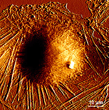
Living fibroblast cell image obtained by AFM

Force mode

In the force operating mode, the tip oscillates vertically while the cantilever deflection is registered.
This method has as its main applications the examination of specific interactions between molecules (antigen-antibody, the complementary strands of DNA), structural interactions (protein folding) or the study of polymer elasticity.


[1] This law states that the lenghtening of materials is directly proportional to the applied force on the same.
[2] Mechanical devices whose function is provide force to move another mechanical device.

Sources: http://www.dme-spm.com/funktion.html
              https://es.wikipedia.org/wiki/Microscopio_de_fuerza_at%C3%B3mica
              http://www.phy.mtu.edu/nue/images/atomicforce/Nanoscope2.jpg
              http://slideplayer.com/slide/6856881/  
              http://www.biozentrum.unibas.ch/research/groups-platforms/overview/unit/lim/


Read more
2.1. Scanning tunneling microscope (STM)

0 comments
In the post titled “Nanotechonology. Introduction” I mentioned that the development of the scanning tunneling microscope played a fundamental role in the progress of nanotechnology, so let’s learn a little more about it and how it works.
The scanning tunneling microscope (STM) was developed by Gerd Binning and Heinrich Rohrer in 1981 at IBM laboratories in Zurich (Switzerland), for which they were awarded the 1986 Nobel Prize in Physics.
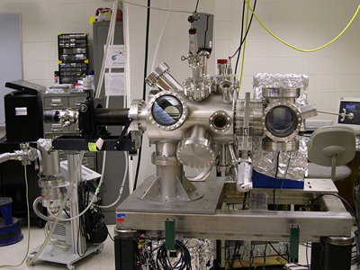
Scanning tunneling microscope
Both the STM and the AFM (atomic force microscope) are classified as scanning probe microscopy instruments, but the first is far more powerful and is able to handle and observe atoms and molecules with higher resolution.

The STM is used to take images of conductive surfaces at an atomic scale of around 2 Å (2*10-10 m). It can also modify the examined sample by manipulating individual atoms, setting off chemical reactions and originating ions by replacing electrons from certain atoms with others.

The scanning tunneling microscope is a non-optical microscope (it does not use rays of light to take us down to nano-dimensions) whose operation is based on quantum mechanic principles. An extremely fine probe is positioned over the specimen under study at a distance of the diameter of an atom, applying a voltage between them. Depending on the characteristics of that voltage, the electrons can jump from one side to the other by tunneling effect[1], producing a weak current flow, known as “tunneling current”, whose value is approximately a few picoamperes (1 pA = 10-12 A).
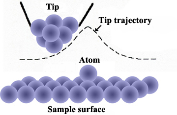
Trajectory of the probe tip over the sample
The stylus probe is very sharp, with a tip formed by a single atom. It scans the surface at a very slow speed, raising and lowering (by a piezoelectric mechanism[2] to control its level) in order to maintain a constant distance and signal, which permits the inspection of the tiniest detail of the sample that is being scanned.
The vertical movement of the stylus is registered allowing the inspection of the surface structure atom by atom, producing a contour map of the surface generated by a computer.

Electrical insulating materials cannot be analysed by this technique because, as their electrical charges do not flow freely it consequently makes it impossible to conduct any kind of current between them and the probe tip.

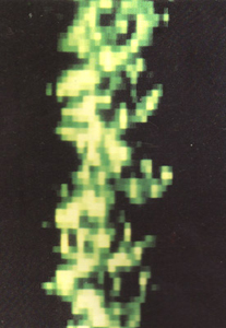
Parallel double-stranded DNA. Image obtained by STM
Although these instruments show an optimal operation examining conductive materials, they can also provide topographical characterisations of organic molecules such as DNA or proteins.

This type of microscope plays an important role in physics, specifically in the study of  semiconductor surfaces and in the field of microelectronics. It also proves to be important in chemistry, studying superficial reactions like catalysis or in nanoscale chemistry laboratories, where the analysis of the physical structure of synthetic chemical compounds and material defects is essential.


[1] The tunnel effect consists in those quantum particles (electrons in this case) that in spite of the fact of not having enough kinetic energy (energy associated with body motion), they penetrate and cross a space that, in principle, would be impossible, due to the presence of a potential barrier that should block their flow.
[2] Materials that are deformed through expansion or contraction, under the action of an electric field.

Sources: http://education.mrsec.wisc.edu/130.htm              
              http://www.nobelprize.org/educational/physics/microscopes/scanning/
              http://hoffman.physics.harvard.edu/research/STMintro.php
              http://www.uwec.edu/Matsci/center/instrumentation/


Read more

1.3. Basic and applied science

0 comments
For a long time, and even now, the value of science has been a topic of debate. This debate has broadly divided science into two categories according to its goals, but not necessarily 'value':

      Basic science or pure science: the search for knowledge regardless of its possible applications in the short run. Its immediate goal is knowledge just for knowledge's sake, no matter whether there is a practical application or not.

      Applied science or technology: whose objective, on the contrary, is to enable solutions to everyday problems, which, as a general rule, are defined by researchers.


Basic science. What value?

There are some people who define applied sciences as “useful” and basic sciences, however, as “useless” and therefore deserving of less attention. However, if we have a look at the history of science, basic knowledge has enabled, subsequently, the development of remarkable applied science. I will list examples below:

Applied scientific discoveries at the famous CERN (European Nuclear Research Centre) initially can be seen as purely theoretical, but contrary to what most people think, these discoveries have day to day applications like the Internet. For example, discoveries at CERN allowed the development of the world's first web site. Also within the computing world, CERN has made feasible the development of GRID, which is a distributed computing system capable of managing the 15 million GB of data that the CERN generates each year. This GRID system allows the distribution of this amount of information and its access by researchers all over the world.

In the medical field, the development of particle accelerators like LHC (Large Hadron Collider) at CERN has provided the emergence of techniques for the treatment of certain diseases like cancer.
One example of these practical applications is hadrontherapy where the tumour is bombed by protons, which enable both more accurate cancer therapy as well as one with reduced patient side effects. Great progress has also been achieved in noninvasive diagnostic imaging techniques like PET (positron emission tomography).

The environment benefits from the advances accomplished at CERN as well, since the complex electronic systems used for detecting particles in accelerators can be applied in environments in which there is the risk of a radiation leak like nuclear power plants in order to enable early detection.
The former accelerator to LHC, LEP, was built using plastic that did not contain sulphur or halogen compounds, which in case of fire, did not produce extremely toxic fumes. These new non-toxic plastics have now been adopted extensively by industry.

Whilst it is arguable that disproportionate amounts of money are spent in order to enable these discoveries, nonetheless I believe that a large number of solutions could not be found without a broad theoretical knowledge produced by basic science.

Source: OpenStax College, Biology. OpenStax College. 30 May 2013.
            http://elgrancolisionadordehadroneshoy.blogspot.com.es/2014/06/aplicaciones-practicas-posibles-riesgos.html
            http://cern123.galeon.com/Beneficios.html             


Read more

1.2. The scientific method

0 comments
The scientific process normally begins with an observation (usually a problem that needs a solution) which leads to a question. Researchers, from this question, are going to follow the next steps:

      Proposing a hypothesis
Let’s remember that a hypothesis is a suggested explanation that can be tested. As several hypothesis can be capable of answering a single question (as we saw previously), several hypothesis can be proposed to solve a single problem. Once a hypothesis has been chosen, a prediction can be formulated with the following structure:  “If . . . then . . .”

    ▣      Hypothesis verification
Due to the fact that natural phenomena are not always as usual and flexible as we would like, observation is not often enough and researchers must carry out one or more experiments to get rid of one or more of the initial hypothesis.
Each experiment consists of:
      At least one variable which is any part of the experiment that can change during the same.
      At least one control group that contains the same features as the experimental group but they are not applied the hypothesis in study. By modifying variables, we can discover how they affect the experimental process and if the results of the experimental group are different from the control group such a difference is due to the hypothesis we are testing instead of external factors.

We can affirm, without a doubt, that the Italian physicist Galileo Galilei was the first one introducing experimentation in a systematic way in the world of natural sciences. Without experimentation, modern science would never have accomplished the advances we have today, that is why laboratories have become so essential for researchers.

Science, unlike general thinking, does not intend to prove anything because scientific knowledge, as time goes by, changes with new acquired knowledge. The objective is, hence, to test if the proposed hypothesis are refuted or disproved, which is general known as “falsifiable”.

The several steps in the scientific method can be represented as follows:
To clear up ideas I will give you a simple example:
Let's imagine you want to watch your favourite TV show but the television does not turn on (observation), so you wonder why (question).
The first idea you think of is there might have been a power cut (hypothesis), to check you do the following “experiment”: you switch on the lamps in your dining room and they light on (analyse the results), so the hypothesis is incorrect.
Then, you decide to look if your TV monitor is unplugged (second hypothesis), you do it (experiment) but the TV is plugged into the mains (analyse the results), this second hypothesis is also incorrect.
Thirdly, you check if the batteries in your remote control are used up (third hypothesis) and the “experiment” you do is to replace them with new ones, finally you manage to turn on the telly (analyse the results), so this last hypothesis was the correct one.

No doubt, this is a rather simplistic example of our daily life but allows us to know how science works with much more complex, significant and tough problems.

Source: OpenStax College, Biology. OpenStax College. 30 May 2013.


Read more

1.1. What is science? How it works

0 comments
Science from the latin “scientia” (knowledge) can be defined as the knowledge that covers general trues or the way general laws work, especially developed and examined through the scientific method. Therefore, the scientific method plays a fundamental role in science and is a research method with very well defined steps, among them experimentation and observation.  

One of the most important aspects in this method is to check the proposed hypothesis through repeatable experiments, being a hypothesis a possible explanation about a fact that can be tested. At the same time, a hypothesis could become a verified theory, which are examined and verified through phenomena or observations. It should be pointed out that a well proposed hypothesis does not imply its validity, because that hypothesis could be false.

Let’s imagine, in a similar way like Galileo supposedly did, we are on the roof of a building and we would like to measure how long it takes a ball to get to the ground, we can formulate the following hypothesis:

      This time depends on what material the ball is made of.

      This time period is determined by the building height.

      This time interval depends on the ball mass.

      Finally, this time is subject to the look of the ball.

    The first, second and third hypothesis can be true or false but they will always be verified designing several experiments. For instance, we can use balls made of diverse materials (iron, wood, plastic…), with different masses (50 g., 150 g.,    250 g…) or we could throw it from buildings with different heights. However, the last hypothesis is not valid, because appearance cannot be measured and therefore, cannot be tested in experiments.

    The common goal in all sciences is to know. Scientists search to understand the world and the way it works, to do it they use two logical reasoning methods:

          Inductive reasoning: a logical reasoning that uses observations related to each other in order to achieve a general conclusion. From lots of observations, raw data (qualitative and quantitative) and an in-depth analysis, scientists deduce conclusions (inductions) based on evidences.

          Deductive reasoning: this kind of reasoning uses general principles or laws to predict specific results. In consequence, it is a thinking pattern that moves in the opposite direction to the former one. Starting from general principles, a scientist can extrapolate particular results which are valid as long as those general principles are also correct.

    Both types of reasoning are used to allow the advance of scientific knowledge.

    Fuente: OpenStax College, Biology. OpenStax College. 30 May 2013.


    Read more