The endomembrane system is a group of organelles and membranes that work together to modify, pack and transport lipids and proteins. This system includes the nuclear envelope, lysosomes and vesicles that we have already mentioned in the previous post: “Prokaryotic and eukaryotic cells. Main components” , the Golgi apparatus and the endoplasmic reticulum.
The endoplasmic reticulum (ER)
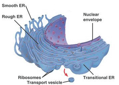
The endoplasmic reticulum is made up of a set of membranous sacs and interconnected tubules that function colectively to modify proteins and synthesise lipids. These two functions are carried out in two different areas: the rough endoplasmic reticulum (RER) and the smooth endoplasmic reticulum (SER).
Rough endoplasmic reticulum (RER)
The rough endoplasmic reticulum receives this name because of the large number of ribosomes that are stuck to its cytoplasmic surface.
The ribosomes transfer the proteins that they synthesise to the lumen of the RER where they undergo modifications and then they are incorporated into the plasma membrane or they are secreted outside the cell through vesicles that bud from the RER membrane.
Enzymes, hormones and even some phospholipids generated by the RER undergo these processes to become part of the cell membranes.
The rough endoplasmic reticulum is mainly found in cells that secrete a large amount of proteins, like hepatic cells.
Smooth endoplasmic reticulum (SER)
The smooth endoplasmic reticulum is next to the RER but, unlike this one, it has few or no ribosomes stuck to its surface.
Among its functions we can find the synthesis of lipids, proteins, steroid hormones, detoxification of poisons and drugs and the storage of calcium ions.
In the muscle cells, the smooth endoplasmic reticulum is called the sarcoplasmic reticulum (SR) and the SR is in charge of accumulating the necessary calcium ions for muscle contraction.
The Golgi apparatus
Before reaching their destination, the proteins and lipids that are carried by the vesicles of the ER must be tagged, classified, packed and distributed. The Golgi apparatus, which consists of a series of flattened membranes, performs these functions.
The receiving side of the Golgi apparatus is called the cis face, whereas the opposite side is called the trans face.
The receiving side of the Golgi apparatus is called the cis face, whereas the opposite side is called the trans face.
As lipids and proteins travel through the Golgi apparatus they undergo a set of modifications that enable classification; the most common modification is the addition of short chains of sugar molecules.
Afterwards, they are tagged with phosphate groups or other small molecules so that they can be delivered to their appropiate destinations.
Finally, they are packed into secretory vesicles that emerge from the trans face of the Golgi apparatus. Some of these lipids and proteins are placed in other parts of the cell, whereas others are fuse with the plasma membrane and they release their content outside the cell.
Immune system cells that secrete antibodies are characterised by having a large amount of Golgi.
Lysosomes
In addition to their digestive role and recycling of organelles, lysosomes are an important component of the endomembrane system too.
Lysosomes use their hydrolytic enzymes to destroy those pathogens that penetrate into the cell.
Lysosomes are used by a special type of white cell called macrophage. These cells via phagocytosis or endocytosis invaginate (fold) their plasma membranes to surround and enclose the pathogen. Later, these pathogens are destroyed under the action of the hydrolitic enzymes of lysosomes.
THE CYTOSKELETON
The cytoskeleton is the group of protein fibres that maintain the cell shape, attach the organelles in their appropiate positions, enable the vesicles to move in the cell and allow the cells of multicellular organisms to move.
Microfilaments
Of the three kinds of protein fibres of the cytoskeleton, microfilaments are the narrowest with a diameter around 7 nm.
They are two intertwined fibres of the actin protein, consequently they are also known as actin filaments.
They participate in processes that require movement such as the cell division in animal cells. They provide rigidity and shape to the cell.
They are two intertwined fibres of the actin protein, consequently they are also known as actin filaments.
They participate in processes that require movement such as the cell division in animal cells. They provide rigidity and shape to the cell.
Intermediate filaments
They purely have a structural function; they bear strain and fix the nucleus and other organelles in the necessary locations.
Their diameter is between 8 and 10 nm and they are made of several twisted protein fibres.
Within this category, keratin filaments are the best known; they strengthen nails, skin epidermis and hair.
Their diameter is between 8 and 10 nm and they are made of several twisted protein fibres.
Within this category, keratin filaments are the best known; they strengthen nails, skin epidermis and hair.
Microtubules
Among the most important roles performed by microtubules, we find the movement of the replicated chromosomes to opposite ends of the cell during the cell division. They also provide a path for vesicles to move inside the cell and help the cell to maintain compression.
They are the widest components of the cytoskeleton with a diameter of around 25 nm.
They are the widest components of the cytoskeleton with a diameter of around 25 nm.
Flagella and cilia
Flagella are moving appendages that extend from the cell membrane making possible the movement of those cells that have them (e.g., sperm).
On the contrary, cilia are short and are found widespread along the surface of the plasma membrane.
Like flagella, they enable the movement of cells (paramecia) or substances over the cell surface, like for example the cilia of the Fallopian tubes that move the ovule towards the uterus.
Like flagella, they enable the movement of cells (paramecia) or substances over the cell surface, like for example the cilia of the Fallopian tubes that move the ovule towards the uterus.
CELL CONNECTIONS
As you can guess, if cells have to work together they must communicate with each other. Let’s see what methods they use to achieve it.
Extracellular matrix of animal cells
The major role of the extracellular matrix is to hold cells together to form a tissue and to allow cell communication within that tissue.
The extracellular matrix is primarily made up of a sort of protein called collagen, intertwined with other types of proteins that contain carbohydrates (proteoglycans).
The extracellular matrix is primarily made up of a sort of protein called collagen, intertwined with other types of proteins that contain carbohydrates (proteoglycans).
Overall, the cell communication is performed as follows:
Cells have quite a few receptors on the surface of their cell membranes.
When a molecule within the matrix joins the receptor, it modifies the molecular structure of the receptor. The receptor, in turn, changes the arrangement of the microfilaments located within the membrane. These changes produce chemical signals that reach the nucleus and they activate and deactivate the transcription of specific sections of DNA, which influences the creation of the related proteins.
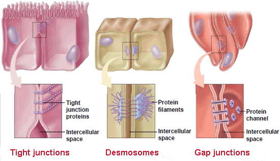
Cells can also communicate through direct contact via intercellular joints, which are formed by diverse kinds of proteins. In animal cells, there are three categories of these joints:
▣ Tight junctions: they seal the plasma membranes of adjacent cells creating a waterproof barrier between them. Their main components are occludin and claudin proteins. These tight junctions are found, for instance, linking the epithelial cells of the urinary bladder.
▣ Desmosomes: they work like spot welds between the epithelial cells of organs and tissues that undergo contraction such as the skin, the heart and muscle cells. Desmosomes are composed of short proteins named cadherins.
▣ Gap junctions: they act like pores and channels that enable the transport of ions and nutrients. They play a significant role in the cardiac muscle, where they allow the movement of the electrical signal that contracts this muscle.
They are made up of a group of six proteins called connexins, which are arranged in the cell membrane in a donut-like configuration known as connexon.
They are made up of a group of six proteins called connexins, which are arranged in the cell membrane in a donut-like configuration known as connexon.
Sources: OpenStax College, Biology. OpenStax College. 30 May 2013.
http://www.oncoursesystems.com/images/user/9341/10845583/rough%20er.bmp
http://apocketmerlin.tumblr.com/post/14923100822/a-summary-of-the-functions-of-major-eukaryotic
http://iesicaria.xtec.cat/~SBG/BiologiaCurtis/Seccion%201/1%20-%20Capitulo%205.htm
https://ohhaitrish.wordpress.com/2012/02/12/unit-one-compilation/


Your opinion matters