• Biology

    Biology

    The atom, biological macromolecules (carbohydrates, lípids, proteins, nucleic acids), cell biology, cancer

  • Drugs

    Drugs

    Drug development, pharmacodynamics, pharmacokinetics, toxicology

  • Nanomedicine

    Nanomedicine

    Introduction to nanotechnology, diagnostic devices, drug delivery, regenerative medicine

  • Miscellaneous

    Miscellaneous

    A more in-depth explanation of topics previously mentioned in the other sections

Posts for label: Biology

Posts for label: Biology

4.5.2. Cell reproduction II

0 comments
The cell cycle is a set of ordered events that involve the cell growth and division to produce two new daughters cells.

Prior to cell division, cells go through a series of stages of growth, DNA replication and division, carefully regulated, with the objective of creating two identical cells.

The two main phases of the cell cycle are the interphase and the mitotic phase. During the first stage, the cell grows and replicates its DNA. While, in the mitotic phase, the replicated DNA and the cytoplasmatic content come apart and the cell divides.


Interphase

The interphase is the longest period of the cell cycle, it occurs between two mitosis and we can distinguish the following phases within it:
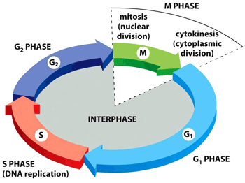
Interphase stages
      G1: the cell grows, reaching its critical size. RNA and proteins are synthesised and the centrosome is duplicated. If conditions are appropiate, the cell passes the restriction point (a G1 phase checkpoint) and the next step begins.
Differentiated cells that do not divide (e.g., neurons or cells from the cardiac muscle) leave the cell cycle at this point and start the G0 phase[1].
      S: DNA is replicated. In this way, two daughter cells inherit the same genome from the mother cell.
      G2: repair phase of DNA and preparation for mitosis. The cell doubles its size compared to G1 phase.


Mitotic phase

The mitotic phase is a multistep process in which the duplicated chromosomes align, come apart and move to the two new daughters created.
The first stage of the mitotic phase is called karyokinesis, mitosis or nuclear division and the second step is known as cytokinesis, which is the physical separation of the cytoplasmatic components into the two daughter cells.
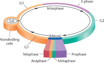
Mitosis, where the genetic material is divided equally, comprises the following stages:
Somatic cell cycle
      Prophase: chromatin is condensed, the mitotic spindle[2] is formed and the nucleolus disappears.
      Prometaphase: the nuclear envelope is removed and the chromosomes join the microtubules through the kinetochores[3].
      Metaphase: the chromosomes align randomly in a plane called metaphase plate, or equatorial plane, between the two poles of the cell.
      Anaphase: the microtubules of the spindle become shorter, which causes the separation of the two sister chromatids.
      Telophase: the chromosomes decondense and the nuclear envelope, along with the nucleoulus is formed. Lastly, the mitotic spindle disappears.

During the last step of the mitotic phase, cytokinesis, a contractil ring of actin filaments is produced which divides the cytoplasm into the two daughter cells.
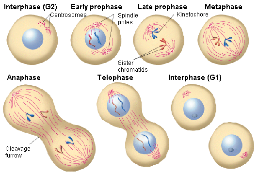
Mitotic phase stages


Control of the cell cycle

The duration of the cell cycle is higly variable, even in cells of the same organism. In human beings, it ranges from just a few hours in the embryonic cells, to two - five days for epithelial cells, to a whole human life for specialised cells, like cardiac muscle cells or cortical neurons.

The time that a cell spends in each phase of the cycle varies too. Thus, for example, those human cells with a 24-hour cycle spend approximately nine hours in the G1 phase, ten hours in the S phase, around four and a half hours in the G2 phase and about 30 minutes in the M phase.

Each step of the cell cycle is controlled by mechanisms both internal and external to the cell.

Regulation of the cell cycle by external mechanisms
Both the start and the termination of the cell replication cycle are activated by events external to the cell.
For instance, events such as the death of a near cell, the release of hormones that favour growth or the cell reaching a specific size can start cell division.
Regardless of the type of externally received message, a series of internal effects then lead it to the interphase.
From this starting point, each required parameter in each phase must be satisfied so that the cell cycle can continue.

Regulation at internal checkpoints
The checkpoints of the cell cycle prevent mistakes in the duplication or in the distribution of chromosomes (which can cause mutations).
We can distinguish three checkpoints:
      The G1 checkpoint: also named the ‘rectriction point’ in mammals. It acts at the end of G1 phase and determines if all the necessary requirements are fulfilled to proceed the cell division.
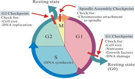
Internal checkpointsSi no se cumplen, la célula puede parar el ciclo hasta que las condiciones adversas se solucionen o puede pasar a la fase G0 y esperar señales que indiquen que las condiciones han mejorado.
      The G2 checkpoint: carries out its role during the transition between G2 to M phases. Its most important function is to make sure that the chromosomes have been replicated and that replication has been accomplished without DNA damage. If damage is detected, the cycle stops and the cell tries to complete the DNA replication or repair the damaged one.
      The M checkpoint: is also known as the spindle checkpoint. It acts near the end of the metaphase stage of karyokinesis. It determines whether the sister chromatids are firmly and correctly bound to the spindle microtubules by the kinetochores.

The control system of the cell cycle could be compared to the program of a washing machine, where the washing machine only carries out its functions when the signals from its sensors (lock door, levels of water and detergent, power supply) indicate that it is ok to move forward during the diverse stages of the washing cycle.


Regulator molecules of the cell cycle

In addition to the checkpoints, there are some intracellular molecules that regulate the cell cycle and can act individually or affect the activity or production of other regulator molecules.
Regulator molecules are classified into two groups:
Regulator molecules of the cell cycle (cyclins and CDKs)
      Those molecules that promote the progress of a cell to the next phase (positive regulation). They are, in turn, divided into two types: cyclins[4] and cyclin-dependent kinases (CDKs)[5].
      The second group are negative regulators, which are in charge of halting the cell cycle. The best-known negative regulators are retinoblastoma protein (Rb), p53 and p21.
Retinoblastoma protein is a suppressor protein for tumours that is altered in many kinds of cancer (prostate, breast), but it is retina cancer from where it takes its name.
P21 inhibits the cell cycle, with its levels controlled, in turn, by p53, which regulates cell growth and controls DNA damage. This protein can halt the cycle if needed, and is able to cause apoptosis too.


[1] Permanent or temporary quiescence (inactivity) phase in which the cell cycle stops.
[2] Set of microtubules whose function is to enable the migration and correct separation of the chromosomes during mitosis.
[3] Protein structures whose function it is to initiate, control and supervise the movement of the chromosomes during cell division.
[4] Proteins synthesised during the interphase and destroyed at the end of the mitosis. They regulate the enzymatic activity of CDKs.
[5] Enzymes that activate or inhibit other proteins by phosphorylating them (adding phosphate groups).

Sources: OpenStax College, Biology. OpenStax College. 30 May 2013.
              http://www.biocancer.com/journal/1219/41-regulacion-positiva-del-ciclo-celular
              http://www.pucmmsti.edu.do/websise/estudiante/materias/201120122/ST-BIO-112-T-003/04.%20Regulacion%20del%20ciclo%
              20celular%20Apoptosis%20Cancer%20%20.pdf
              http://bio3400.nicerweb.com/Locked/media/ch02/interphase.html
              http://www.slideshare.net/obessionforu/cell-cycle-and-molecular-basis-of-cancer
              http://biologicalexceptions.blogspot.com.es/2013_01_01_archive.html
              http://gleesonbiology.pbworks.com/w/page/7537859/C9
              http://www.bioscirep.org/content/30/4/243


Read more

4.5.1. Cell reproduction I

0 comments
Human beings, like other organisms that reproduce sexually, start life from a zygote or fertilised egg.
Later, trillions of cell divisions take place in a controlled way to lead to a complex multicellular human being, or in other words, a single cell is the common ancestor of the rest of the organism’s cells.
Even when a living being has developed completely, cell division is still necessary for the development, maintenance and repair of cells and tissues. This division is strictly regulated to avoid consequences that might be fatal for that organism.


Cell division

Cell reproduction via the cell cycle constitutes the foundation that enables the continuity of life from one cell to another. This cycle, strongly regulated, includes the diverse stages of a cell life from the division of a single parent cell to the reproduction of two new daughter cells.

Before going on with the cell reproduction process, it is necessary to understand the function and structure of the genetic information of a cell.Chromosome structure
The number of chromosomes[1] of eukaryotic cells varies depending on the species.
In human beings, body cells (somatic cells) have 46 chromosomes, while sex cells or gametes (eggs and sperm) have 23 each.
Somatic cells have two sets of chromosomes (2n[2]) in a configuration called diploid.
Gametes, that have a single set of chromosomes, are designated as 1n and they are known as haploid cells.

Genes (functional units of the chromosome) determine particular features by coding specific proteins, to represent variations in specific features. For instance, eye colour is a characteristic whose traits can be brown, blue or green.
These traits in an individual are established by the inherited genes from each parent.

If the DNA of those 46 chromosomes in a human cell unfolded from one end to the other, it would measure around 2 metres and its diameter just 2 nm. Taking into account that the typical size of a cell is about 10 μm, DNA has to condense itself to fit into the cell nucleus and must be quickly accesible so that genes can be expressed.

During several phases of the cell cycle, the long chains of DNA are highly condensed in the chromosome. There are a number mechanisms by which chromosomes are compacted, in all of them various classes of proteins help to organise and pack the chromosomal DNA.

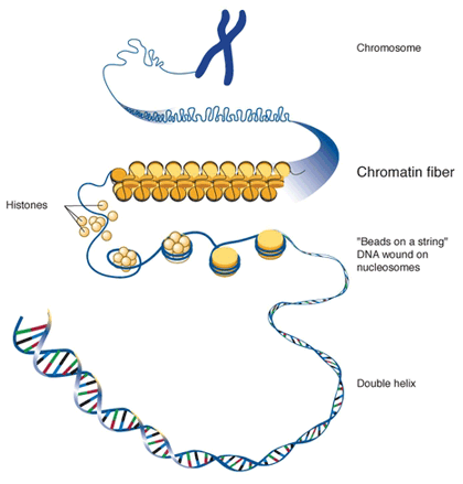
In the first level of compaction, DNA is strongly packed around a core of eight structural proteins (histones) in regular intervals along the chromosome, forming the structure known as chromatin.
The fundamental unit of organisation for chromatin in eukaryotic cells is the nucleosome, composed of a DNA fragments around 200 base pairs (bp) long and a histone octamer (group of eight). This structure compacts seven times the DNA molecule which under a microscope looks like a ‘necklace of beads’ with a 10 nm diameter.
DNA condensation levels

In the second level of compaction, the nucleosome fibres and the linker DNA[3] roll into 30 nm fibres (DNA molecule is now 50 times packed).

The third level of compaction uses fibrous proteins to compact chromatin, creating radial loops of 250-400 nm long. These proteins make sure that each chromosome in the non-dividing phase occupies a certain area in the nucleus avoiding overlap.

In higher levels of organization we find chromatids (700 nm - 1μm long). When two of these chromatids join, through a region called the centromere, they form metaphasic chromosomes[4].


[1] Highly organized structures comprising DNA and proteins, which contain most of the genetic information of an individual.
[2] n indicates the number of chromosome sets.
[3] DNA regions that are not transcribed and are located between genes.
[4] Metaphase: second phase of mitosis (cell division) in which the nuclear membrane is removed and the chromosomes are placed in the equator of the cell.

Sources: OpenStax College, Biology. OpenStax College. 30 May 2013.
              https://www.quora.com/What-are-the-differences-between-chromosomes-chromatids-and-chromatin
              https://burningscience.wordpress.com/biology/dna-structure-and-replication/
              http://27.109.7.67:1111/econtent/cell-communication/principles-part-3.php
              https://www.quora.com/What-are-the-differences-between-chromosomes-chromatids-and-chromatin


Read more
4.4. Cell communication

0 comments
In the same way that social organisations require communication between individuals and their environment to maintain societal cohesion, cells must also be able to interact with their environment.

In order to respond to external stimuli, cells have developed complex communication systems that allow them to receive messages, transfer information across the plasma membrane and produce changes within the cell in response to those messages.

Cells of multicellular organisms constantly send and receive messages to coordinate the actions of cells, tissues and distant organs. The capacity to send messages in an effective and quick way allows cells to coordinate and adjust their functions.
This capacity to communicate via chemical signals, initially developed by individual cells, was a vital attribute for the evolution of multicellular organisms.


Signalling molecules and cellular receptors

Cells are in contact via intercellular signaling (communication between cells) and intracellular signaling (communication within the cell).
Signalling cells are responsible for secreting ligands[1] or they bind to target cells, triggering a event chain within them.

In multicellular organisms, chemical signalling is classified into four categories: endocrine signalling, autocrine signalling, paracrine signalling and direct signalling by gap junctions (juxtacrine signalling).
Endocrine signalling is performed by hormones across long distances through the bloodstream.
In autocrine signalling, signals are received by the same cell that sends them or other close cells of the same type.
Paracrine signalling also acts at short distances under the action of ligands that travel through the liquid medium of the extracellular matrix.
Lastly, gap junctions enable signalling molecules to flow between neighbouring cells.
Types of cell signalling

Types of receptors

Receptors are proteins located within, or on the surface of target cells that bind to ligands. Receptors are thus divided into two different classes: internal receptors and cell-surface receptors.

Internal receptors are located in the cell cytoplasm attached to the ligand molecules that pass through the cell membrane. These receptor-ligand complexes move to the nucleus and there they interact directly with the cellular DNA.

Cell-surface receptors convey a signal from outside the cell to the cytoplasm. They comprise three components: a region outside the cell to which the ligand binds (also called extracellular domain), a hydrophobic central region in the membrane and the intracellular domain within the cell.
They are divided into three categories:
      Ion channel-linked receptors[2]. When they bind to their specific ligands they form a channel across the plasma membrane through which certain ions can pass.
      G-protein-linked receptors. They interact with G-proteins[3] on the cytoplasmatic side of the cell membrane.
Once the G-protein binds to the receptor, the resultant compound activates the G-protein, which releases GDP[4] and pick up GTP[5], interacting with other enzymes or ionic channels to transmit the signal.
      Enzyme-linked receptors. They carry a signal of membrane-bound enzymes from outside the cell to the intracellular domain. The union with the ligand causes the enzyme activation.
Types of cell receptors
Small hydrophobic ligands (e.g. steroids) are able to penetrate the cell membrane and stick to internal receptors.
On the other hand, hydrophilic receptors (soluble in water) are unable to pass through the membrane; consequently they link with cell-surface receptors, which convey signals inside the cell.


Propagation of the signal

The binding of a ligand to a receptor enables signal transduction[6] throughout the cell.
The chain of events that creates signal transmission is called the ‘signalling cascade’ or ‘signalling pathway’. This pathway can be very complex as a consequence of the interaction among different proteins.
One of the most important events in this process is the phosphorylation of molecules by way of specific enzymes known as kinases[7].
Protein phosphorylation processPhosphorylation adds a phosphate group to residues (R-groups) of the following amino acids: serine, threonine and tyrosine, activating and deactivating the protein to these amino acids belong to or changing its shape. Also, small molecules such as nucleotides can be phosphorylated.

Once a receptor has been activated, the signal propagates throughout the cell via the cytoplasm by modifying the behaviour of certain cellular proteins. These signals are released by small non-protein molecules, called second messengers. Among them are: cyclic AMP (cAMP), calcium ions (Ca2+), inositol triphosphate (IP3) and diacylglycerol.


Response to the signal

The initiation of signalling routes is triggered in response to external stimuli. The effects of these responses are quite diverse, depending on the type of cell involved, as well as internal and external conditions.
Among these effects are cell growth, protein synthesis, changes in the cell metabolism and even cell death.

But these signalling routes have their most significant impact on the cell by initiating gene expression[8].
Thus, some routes activate enzymes that interact with transcription factors of RNA and others regulate protein transduction from mRNA.
Other routes again, however, work on the cellular metabolism, e.g. they enable muscle cells to communicate their functional energy requirements (these requirements are in the form of glucose).

Signalling routes play a very significant role in cellular growth too, which is stimulated by external signals, called ‘growth factors’ (types of ligands that bind to cell-surface receptors).
Uncontrolled cell growth lead to cancer. Cancer’s origin is often due to mutations presented by those genes responsible for encoding the proteins that are part of signalling pathways.

When a cell is damaged, is unnecessary or is potentially dangerous, the organism must initiate the process that triggers programmed cell death or apoptosis. Apoptosis allows a cell to die in a controlled way, avoiding the release of potentially harmful molecules, in contrast to uncontrolled death or necrosis.
Necrosis and apoptosis processes
Cell signalling controls the process of apoptosis so that the dismantling of cells is carried out in an organised way, as well as the efficiently recycling the different components of dead cells.

The end of the signalling cascade is very important in ensuring that the signal response is appropriate both in time and intensity.
Two of the most common ways of ending signalling within cells is with the degradation of signalling molecules and dephosphorylation of intermediate products of the pathway (previously phosphorylated) via phosphatase[9] enzyme.

Some of the abnormal signals originated in tumour cells are good evidence that termination of cellular cascades at the appropriate time is as important as the timing of initiation.


[1] Small volatile or soluble molecules that bind to other specific molecules (receptors) releasing a signal in the process.
[2] Cell-surface receptors in the channels of plasma membranes that open when a ligand sticks to its extracellular domain.
[3] Signal transducers that transmit information from the receptor to effector proteins.
[4] Guanosine diphosphate: dephosphorylation product of GTP.
[5] Guanosine triphosphate is a purine nucleoside triphosphate used in the cell metabolism. 
[6] Transmission of a signal from outside the cell to its inside.
[7] Enzymes that catalyse the transfer of a phosphate group from ATP to another molecule.
[8] Process by which cells transform the encoding information of amino acids into necessary proteins for their development and functioning.
[9] Enzyme in charge of removing the phosphate group of a molecule that has been phosphorylated.

Sources: OpenStax College, Biology. OpenStax College. 30 May 2013.
              http://mol-biol4masters.masters.grkraj.org/html/Cellular_Signal_Transduction1-Cells_And_Signals.htm 
              http://27.109.7.67:1111/econtent/cell-communication/principles-part-3.php
              http://www.scq.ubc.ca/protein-phosphorylation-a-global-regulator-of-cellular-activity/
              http://medicinembbs.blogspot.com.es/2011/03/programmed-cell-death-apoptosis.html


Read more

4.3.2. Cell respiration II: Oxidative phosphorylation

0 comments
Most of the ATP originated during the aerobic catabolism of glucose does not come from the two previous routes (Glycolysis and Krebs cycle), but rather from the movement of electrons through a series of electron carriers that undergo redox reactions, which causes the accumulation of hydrogen ions in the mitochondrial matrix.

As a consequence, a concentration gradient is formed in which hydrogen ions diffuse out of the mitochondrial matrix by passing through it with the help of the complex called ATP synthase[1]. The current of hydrogen ions promotes the catalytic action of ATP synthase, which phosphorylates ADP, producing ATP.


Electron transport chain

The electron transport chain is the last component of aerobic respiration and the last stage of glucose metabolism that uses atmospheric oxygen.
This oxygen is used as a final receptor for the electrons that have been removed from the intermediate compounds in glucose catabolism.

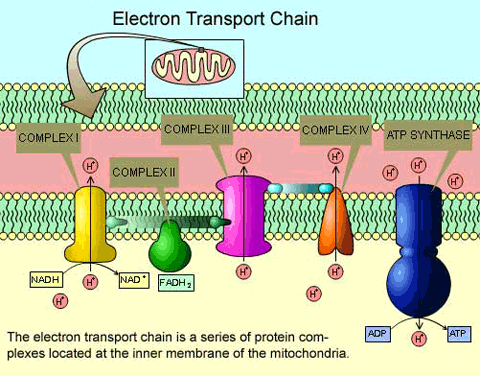
The electron transport chain consists of four large multiprotein complexes (called complexes I-IV) embedded in the inner mitochondrial membrane and two small electron transporters that move the electrons between them.

Electrons participate in a series of redox reactions in which free energy is used to shuttle hydrogen ions across the mitochondrial membrane using the ATP synthase enzyme. This process contributes to the gradient used in chemiosmosis (a mechanism based on the diffusion of ions across a membrane) that produces 90% of ATP created during aerobic glucose metabolism.
Elements of the electron transport chain

The electrons that take part in the transport chain lose their energy gradually, so to complete it, the contribution of high-energy electrons from the NADH or FADH2 compounds is necessary (generated in the previous process (Krebs cycle).

At the end of this metabolic pathway, electrons reduce oxygen molecules to oxygen ions and the extra electrons of these ions attract hydrogen ions (protons) from the surrounding environment, resulting in the release of water molecules and ATP as the final products of the electron transport chain.

The number of molecules created varies depending on a number of factors such as the number of hydrogen ions that the complexes of the electron chain pump across the mitochondrial membrane, the transport of electrons or the use of intermediate compounds produced in these routes for other purposes.


Metabolism without oxygen

In aerobic respiration, the final receptor of electrons is the oxygen molecule (O2) and ATP is produced with the assistance of high-energy electrons transported to the transport chain by the molecules NADH or FADH2.
If aerobic respiration does not take place, the NADH compound must be re-oxidased to NAD+ so that it can be reused as an electron carrier, and in this way the glycolytic pathway continues.
Living organisms employ two different mechanisms to achieve this:
      They can use an organic molecule as the final acceptor of electrons to regenerate NAD+ from NADH in a process called fermentation.
One of the most well known fermentation processes is lactic acid fermentation, used by red blood cells in skeletal muscles of mammals when they lack enough oxygen to continue aerobic respiration.
Lactic acid fermentation
      The second option is to use an inorganic molecule instead.
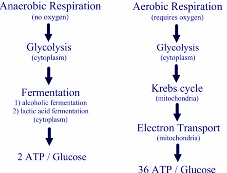
In both, organisms transform energy to use it in the absence of oxygen and they are known as anaerobic cellular respiration.
Aerobic and anaerobic processes


Regulation of cellular respiration

Cellular respiration must be regulated in order to supply the required amounts of energy at any time in the form of ATP.
The cell must regulate, therefore, its metabolism and for that have a broad variety of mechanisms.
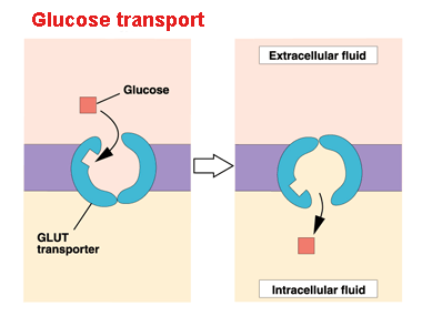
For instance, glucose entering the cell via the plasma membrane is controlled by transport proteins (GLUT proteins). But most of the control of the respiratory process is performed by specific enzymes that act on each route.
Cellular uptake of glucose
These enzymes react to the available levels of the nucleosides ATP, ADP, AMP, NAD+ and FAD, which, in turn, increase or decrease enzyme activity on the routes where they participate.

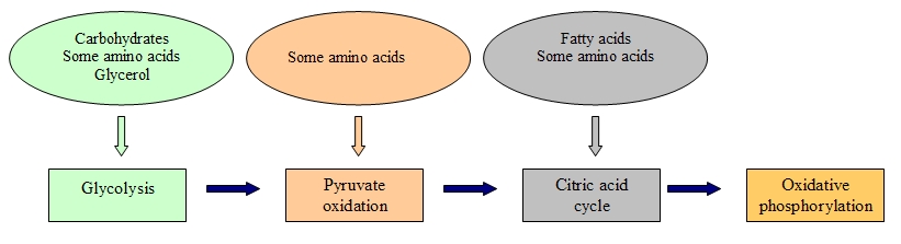
We have seen that glucose metabolism is responsible for providing energy to living cells. Yet, living beings consume a broad range of nutrients other than glucose in their diets. Hence, how do these foods become ATP in our cells?
Eventually the catabolic pathways of lipids, proteins and carbohydrates are connected with glycolysis and citric acid cycle pathways.
These pathways are not closed cycles, but many of their substrates, intermediate and final products are used in other routes.
Connection of carbohydrates, proteins and lipids to glucose metabolism


[1] It is a transmembrane protein complex (enzyme) that catalyses ATP synthesis by the supplied energy from a proton flow (H+) and by adding a phosphate group to ADP.  

Sources: OpenStax College, Biology. OpenStax College. 30 May 2013.
              https://chelseaharripersad.wordpress.com/2013/04/13/the-electron-transport-chain/
              http://healthylifemed.com/aerobic-vs-anaerobic-respiration/
              https://adapaproject.org/bbk_temp/tiki-index.php?page=Leaf%3A+Why+do+cells+need+fermentation+to+continue+glycolysis%3F
              http://163.178.103.176/Fisiologia/general/dinamica/FG05_17a.jpg


Read more

4.3.1. Cell respiration I: Glycolysis. Krebs cycle

0 comments
The energy that enters into our organisms as food is transformed so that cells can use it and perform their functions.
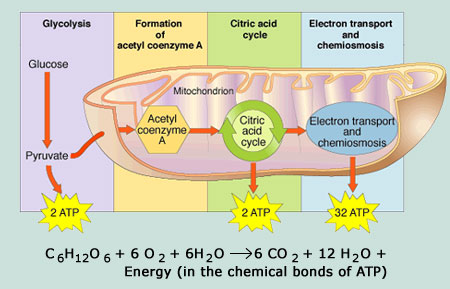
Thus, through a set of metabolic pathways (collectively called cell respiration), the energy of glucose bonds is extracted and turned into an energy form that all living organisms can use.
Metabolic pathways of cell respiration

Energy in living beings

A living cell does not collect excessive amounts of free energy because it could damage and subsequently destroy it. Hence, cells must store that energy in a safe way and free it only when the need arises, which is achieved by the adenosine triphosphate compound (ATP), which is considered the ‘energy currency’ of cells.

ATP allows the cell to store energy for a short time and to transport it within the cell to facilitate endergonic chemical reactions.
ATP’s structure is made up of an adenosine monophosphate molecule (AMP[1]), which consists of an adenine molecule attached to a ribose molecule and one phosphate group. The addition of a second phosphate group produces adenosine diphosphate (ADP) and the addition of a third phosphate group forms finally adenosine triphosphate (ATP).

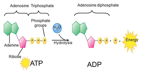
Life to accomplish its processes and obtain energy, it breaks down ATP into ADP[2] constantly, through the hydrolysis ATP reaction, producing, at the same time, an inorganic phosphate ion:

ATP + H2O ADP + inorganic phosphate (Pi)
ATP hydrolysis

The water molecule that takes part in this reaction is split into a hydrogen atom and a hydroxyl group. This water is regenerated when a third phosphate is added to the ADP molecule, which reconstitutes, in turn, ATP. This mechanism works, therefore, like a rechargeable battery.

In practically all organisms, the required energy comes from glucose metabolism. This way, ATP is directly connected to the set of exergonic pathways of the catabolism of glucose and the large number of endergonic pathways that supply energy to cells.

The two processes of ATP regeneration used in combination with glucose catabolism are substrate phosphorylation[3] and oxidative phosphorylation.
In the first mechanism, ATP is produced from ADP and a phosphate group from a reactive.
But most of the generated ATP (90%) during the glucose catabolism is derived from a much more complex process, which ocurrs in the mitochondrion: chemiosmosis. During chemiosmosis in which a proton gradient is involved across the mitochondrial membrane.
The production of ATP using this mechanism is called oxidative phosphorylation because of the involvement of oxygen in it.


Glycolysis

Glycolysis was probably one of the first metabolic pathways used during the evolution of living beings and it is used by nearly all of them.
Glycolysis is the first point of glucose breakdown for the extraction of energy during the cellular metabolism. It does not require oxygen; it is thus an anaerobic mechanism.
Phases of glycolysis pathway
This process is composed of two steps:
      The six carbon atom ring of glucose molecule is divided into two sugar molecules (called pyruvate) with three carbon atoms each one. ATP is needed to cause this separation.
      ATP and high energy electrons are extracted from hydrogen atoms, and are attached to NAD+ compounds (oxidised form of the molecule NAD[4]).
The first stage invests two ATP molecules, while the second stage produces four ATP molecules through the substrate phosphorylation mechanism.
The final outcome is a net gain of two ATP molecules and two NADH[5] for the cell.
In the event that the cell cannot catalyse the pyruvate molecules, it only gets two ATP molecules from a glucose molecule.


Oxidation of pyruvate and the Citric Acid Cycle (Krebs cycle)

If there is oxygen available, aerobic respiration then takes place following the glycolysis process.
Pyruvate molecules originated at the end of that metabolic pathway are transported into mitochondria where cell respiration takes place. There, pyruvate is transformed into an acetyl group, which is collected and activated by a carrier called coenzyme A (CoA)..
The resultant compound, named acetyl coenzime A, is made up of vitamin B5 (pantothenic acid). This compound is used in a wide range of ways by cells, but its principal function is to distribute the acetyl group that comes from the pyruvate to the following stage in glucose metabolism (the citric acid cycle).
Krebs cycle scheme
The conversion of pyruvate to an acetyl group removes a CO2 molecule and two high energy electrons. This step occurs twice, so that the CO2 molecule (2CO2) holds two of the six carbon atoms from the initial molecule of glucose. While the NAD+ molecule picks up the electrons forming NADH that conducts those electrons to later pathways in the production of ATP.

At this point, the glucose molecule that entered the cellular respiration mechanism has been completely oxidised and its potential energy has been transferred to electron transporters or has been used to synthesise a few ATP molecules.

Next, the citric acid cycle also begins in the mitochondrial matrix.
Unlike glycolysis, the cytric acid cycle is a closed cycle, in which the last step regenerates the compound used in the first stage.
The cytric acid cycle is an eight step cycle that comprises a series of oxidation-reduction (redox), dehydration, hydration and decarboxylation reactions that produce: two CO2 molecules, one GTP[6]/ATP molecule and the reduced forms of three NADH molecules and one FADH2[7] molecule.

This cycle is considered an aerobic route because these two last compounds must transfer their electrons to the next pathway of the system, which will use oxygen and will generate ATP.

Some of the intermediate compounds in this cycle can be used to synthesise non-essential amino acids, lipids and sugars that can be energy sources for the metabolic pathways of glucose. Hence, the Krebs cycle is an amphibolic cycle (both anabolic and catabolic).


[1] AMP is one of the nucleotides used in RNA.
[2] Adenosine diphosphate: organic compound made up of adenosine and two phosphate groups.
[3] Addition of a phosphate group to a compound, usually a metabolic intermediate, a protein or ADP.
[4] Nicotinamida adenina dinucleótido: coenzima encontrada en las células vivas, cuya función principal es el intercambio de electrones y protones en las reacciones de producción de energía.
[5] Reduced form of the NAD molecule that therefore, it accepts electrons.
[6] Guanosine triphosphate is another nucleotide required for RNA synthesis and involved in cellular metabolism. Its nitrogenous base is purine guanine.
[7] Reduced form of FAD (flavin adenine dinucleotide: coenzyme that intervenes in metabolic reactions of oxidation-reduction) that accepts two hydrogen atoms.

Fuentes: OpenStax College, Biology. OpenStax College. 30 May 2013.
              http://table4eversquishycells.pbworks.com/w/page/9947625/How%20Cells%20Get%20Energy
              http://bio100.class.uic.edu/lectures/atp_energy.jpg
              https://apbionotebook.wordpress.com/chapter-09-cellular-respiration-fermentation/
              http://msdoranbiology.weebly.com/notes---cp.html


Read more

4.2. Cellular metabolism

0 comments
Practically all tasks performed by living organisms require energy. Energy is not only needed to perform exercise or demanding tasks, but also when we think, and even when we sleep; its use is, therefore, constant.

Energy is used so that immune system cells can absorb, ingest and break down viruses and pathogenic bacteria. It is also fundamental to eliminate toxins and waste, to transport neurotransmitters and hormones and in the synthesis and chemical breakdown of molecules.

Nutrients and other molecules are imported, metabolised (broken down) and synthesised in other molecules, modified in some cases, transported to other cells and probably distributed throughout the body by using energy.

All cellular processes, such as the building and degradation of complex molecules occur through a series of staged chemical reactions.
These chemical reactions within cells, including those that release and use energy, is called cell metabolism.


Metabolic pathways

A metabolic pathway is a series of interconnected chemical reactions that transform one or several substrate molecules gradually, via a group of metabolic intermediates, into a final product or products.

In the case of sugar metabolism, glucose (a simple sugar) is synthesised from smaller molecules:

6CO2 + 6H2 O + energy → C6 H12 O6 + 6O2

While in another different metabolic pathway, glucose is broken down into smaller molecules:

C6H12O6 + 6O2 → 6CO2 + 6H2 O + energy

Hence, metabolism consists of building or anabolism (first reaction) and breakdown or catabolism (second reaction).

These metabolic pathways do not ocurr spontaneously, but each reaction is enabled or catalysed by specific proteins called enzymes, which catalyse both the reactions that release energy and the reactions that require its use.


Potential, free and activation energy

Free and activation energyWhen the bonds in molecules are broken down, they release energy. This is known as potential energy. To carry out their function, cells depend on the extraction of that potential energy.

Free energy is a concept that measures the available energy to perform a task. This free energy is modified during chemical reactions (where energy transfers occur) and this change is denominated as Gibbs free energy (ΔG).
Exergonic and endergonic reactions
The value of ΔG can be negative (whereby energy is released). These reactions are called exergonic or spontaneous. On the contrary, if the value of ΔG is positive then the reaction consumes energy. These reactions are called endergonic or non-spontaneous.

If a reaction is spontaneous, this indicates that its products have less energy than its reactives, and vice versa for non-spontaneous reactions.
Regardless of the sort of reaction, all of them however require an initial contribution of energy to reach the transition state[1]. This contribution is known as activation energy.


ATP: adenosine triphosphate

ATP is the molecule that provides energy to living cells. It is composed of a nucleotide, a pentose (a five carbon atom monosaccharide) and three phosphate groups. The bonds of these three phosphate groups have a high energy content that when are broken, promote a series of reactions and cell processes.
The energy that is released in the ATP hydrolysis reaction:
ATP +H2O → ADP + Pi[2] + energía libre (reacción reversible)
ATP hydrolysis mechanism

Cells use ATP by coupling the exergonic reaction of ATP hydrolysis with endergonic reactions. ATP gives its phosphate group to another molecule during a process known as phosphorylation. This phosphorylated molecule is less stable than its unphosphorylated form, but the added energy after the addition of the phosphate group enables it to perform the necessary endergonic reactions so that the cell can carry out its different functions.


Enzymes

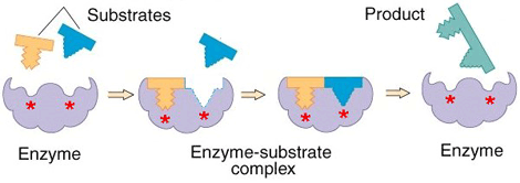
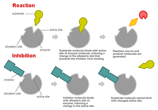
Enzymes (usually one or more polypeptide chain proteins) speed up chemical reactions by diminishing their activation energy requirement.
Enzymes bind to substrates[3] to catalyse reactions in four different ways:
      Taking part directly in chemical reactions by forming transient covalent bonds with substrates.
      Uniting substrates in an optimal orientation.
      Providing optimal conditions so that the reaction can take place.
      Acting on bond structures so that they can be broken more easily.
How an enzyme works

Enzymes are regulated by cell conditions like pH, temperature and their location within the cell (some of them are compartmentalised).
But the most common method by which cells regulate enzymes in metabolic pathways is via feedback inhibition. In this way the products of a metabolic pathway act as inhibitors (normally allosteric[4]) of one or more enzymes involved in the pathway where these products are originated.
Enzyme activity


[1] Very high energy and short duration state that is produced when the reaction reactives approach and experiment a deformation.
[2] Inorganic phosphate group.
[3] Specific chemical reactives on which a specific enzyme performs.
[4] They decrease the enzyme activity by causing structural changes on them, enzyme receptors change and adopt their inactive conformation.

Sources: OpenStax College, Biology. OpenStax College. 30 May 2013.
              http://cienciasdejoseleg.blogspot.com.es/2013/03/plicacion-de-las-variables.html
              http://teenbiotechchallenge.ucdavis.edu/2010_TBC/Peter%20Wang,%20Clara%20Fannjiang,%20William%20Liu/Chemistry.html
              https://mollycools.wordpress.com/2014/03/10/enzymes/
              https://commons.wikimedia.org/wiki/File:Allosteric_competitive_inhibition_3.svg


Read more

4.1.3. Plasma membrane. Cellular transport

0 comments
Among the different functions of the plasma or cell membrane, the most basic is to establish the cell’s limits and keep it functional.
The membrane is selectively permeable. Consequently some elements can enter and exit the cell freely, whereas others need a specialised structure or even energy to accomplish this.

The most significant transport routes that the cell uses through its plasma membrane are:

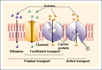
Passive transport

Passive transport is a natural phenomenon where the cell does not need to use any sort of energy to move substances across the plasma membrane.
In passive transport, the substances pass from a high concentration region to a low concentration region.
The various mechanisms of passive transport are:

      Diffusion: a single substance moves from a high concentration area to a lower one until the concentration is equalised on both sides.
A variation of diffusion is the filtration process where materials move through the membrane according to their concentration gradient[1]. At times, the diffusion rate is increased by pressure causing materials to be filtered much more quickly. This is the process that takes place in kidneys where the blood pressure forces big amounts of water and substances dissolved in it (solutes) from the bloodstream to the renal tubules.

      Facilitated transport: diffusion is carried out by the membrane with the help of its embedded proteins, which act like shields for these substances (polar molecules, ions) against the membrane’s repellent forces, facilitating their diffusion inside the cell.

      Osmosis: is a special case of diffusion in which water, not material, is carried across the membrane. Therefore, the membrane limits the diffusion of solutes in water. This process is mediated by a set of specific proteins called aquaporins.
Passive and active transport

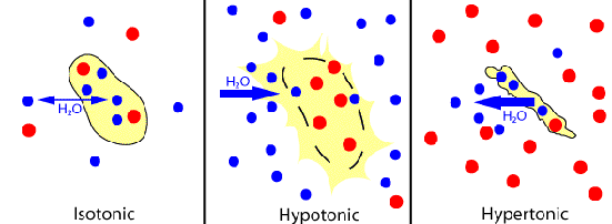
Depending on the relationship between the cell osmolarity[2] and the osmolarity of the surrounding extracellular fluid, we can identify three solution states:

      Hypotonic solutions: the osmolarity of the extracellular fluid is lower than the osmolarity of the fluid inside the cell, therefore water penetrates the cell.

      Hypertonic solutions: the osmolarity of the extracellular fluid is higher than the cytoplasm osmolarity, hence water leaves the cell.

      Isotonic solutions: both the cell and the extracellular fluid have the same osmolarity. As a consequence, there is a constant exchange of water between both regions.
Different types of solutions

Active transport

Active transport, also known as pumps, requires the use of energy in the form of adenosine triphosphate (ATP). This energetic requirement is necessary because the substance is moving against a concentration gradient (i.e., the substance concentration in the cell is higher than in the extracellular fluid or vice versa).
By this mechanism a wide range of different sized molecules can be carried.

In living organisms, there are not only simple concentration gradients but also electrical gradients that indicate a charge difference across the cell membrane.

The cell interior is negative electrically in comparison to the extracellular fluid, which has a higher concentration of K+ (potassium ions) and a lower concentration of Na+ (sodium ions) than the extracellular fluid.
Consequently, the electrical and concentration gradients of Na+ tend to lead these ions into the cell.
The K+ ion situation is more complex; while its electrical gradient also tends to drive these ions towards the interior of the cell, its concentration gradient leads out of the cell.

These combined gradients of electrical charge and concentration that affect an ion are named electrochemical gradient.

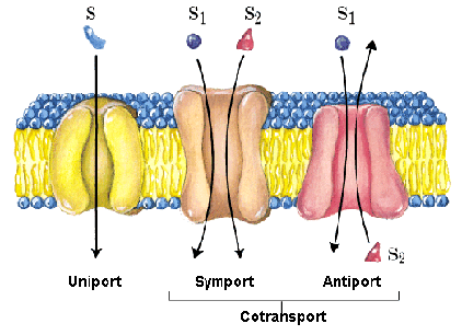
A notable adaptation of the cell membrane for active transport is the presence of specific transport proteins (pumps) that enable the movement of materials on both sides of the membrane. These proteins or transporters are classified into three types:

- Uniporters: they carry a specific molecule or ion in just one direction.
- Symporters: they transport two different ions or molecules in the same direction.
- Antiporters: they also transport two different molecules or ions but in opposite directions.
Classification of transporters

Primary active transport
This type of transport works with the active transport of K and Na and enables secondary active transport to occur at later stage.

One of the most important mechanisms of active transport in animal cells is the Na+ -K+ pump that maintains the electrochemical gradient and the appropiate concentrations of Na+ and K+ in cells.
During this process for every three Na+ ions that are expelled from the cell, two K+ ions penetrate into the cell, therefore the interior of the cell is slightly more negative than its exterior. This charge difference is vital to create the necessary conditions for the second process.

The Na-K pump belongs to the electrogenic pump category, which is a kind of ionic pump that generates a potential electrical charge difference on both sides of the cell membrane.

Secondary active transport (co-transport)
Via this process Na+ ions and other compounds penetrate into the cell taking advantage of the electrochemical gradient created in the previous step.
In this class of active transport a solute that moves against the concentration gradient is co-transported with other solutes that move in favour of its concentration gradient.
Thanks to this method, many diverse molecules, like amino acids or glucose molecules, can enter the cell.
This process is also used to store hydrogen ions of high energy in mitochondria to produce ATP.


Bulk transport

In addition to small ions and molecules, cells must also assimilate and eliminate larger size molecules and particles and in order to achieve it they need a supply of energy. However, these molecules are so large that not even with the supply of energy can they pass through the plasma membrane.
Hence, the cell uses the following mechanisms:

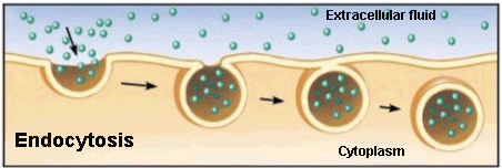
Endocytosis
Endocytosis process
Endocytosis is a type of active transport where particles, such as large molecules, parts of cells and even entire cells are enclosed by invagination of the membrane forming a vesicle whose content is carried from the outside to the inside of the cytoplasm.

Listed are the various endocytosis:

      Phagocytosis: is the process in which cells, or large parts are ‘consumed’ by a cell. This is the method used by a particular type of white cells called neutrophils. These immune system cells surround, cover and destroy the microrganisms that invade our body.

      Pinocytosis: the cell captures the molecules that are needed from the extracellular fluid. The cell membrane invaginates that unwelcome cell, creating a vesicle around the liquid of the external fluid, which is ‘consumed’ and released in the cytoplasm.
For example, the maturation egg process in the uterus is carried out by pynocytosis when the ovule takes in the nutrients that its supply cells release.

      Receptor-mediated endocytosis: uses receptor proteins from the cell membrane that have a specific affinity to binding to particular substances.
Endocytosis in animal cells

Some human diseases are caused by the malfunction of this mechanism. It is the case of familial hypercholesterolemia, in which the receptors of bad cholesterol are faulty or completely absent.

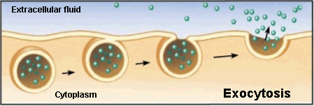
Exocytosis
Exocytosis process
Exocytosis is the opposite to endocytosis in which the cell expels material from its interior to outside.
This waste material is covered with a membranous sac that fuses to the plasma membrane and then opens to release its content into the extracellular space.
The neurotransmitter secretion by vesicles in the synaptic gap of neurons follows this process.


[1] Different concentration of molecules between two regions.
[2] It describes the overall concentration of solute in a solution.

Sources: OpenStax College, Biology. OpenStax College. 30 May 2013.
              http://www.educ.ar/sitios/educar/recursos/ver?id=14378
              http://sevendaysperweek.blogspot.com.es/2015/09/spm-biology-3-movement-of-substances_20.html
              http://cbc.arizona.edu/classes/bioc462/462a/NOTES/LIPIDS/transport.html
              Nelson & Cox, Lehninger Principles of Biochemistry, 3rd ed., 2000
              http://es.slideshare.net/exitoinevitable/clulas-31270712
              https://micro.magnet.fsu.edu/cells/endosomes/endosomes.html


Read more

4.1.2. The endomembrane system. The cytoskeleton

0 comments
The endomembrane system is a group of organelles and membranes that work together to modify, pack and transport lipids and proteins. This system includes the nuclear envelope, lysosomes and vesicles that we have already mentioned in the previous post: “Prokaryotic and eukaryotic cells. Main components” , the Golgi apparatus and the endoplasmic reticulum.

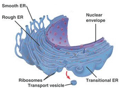
The endoplasmic reticulum (ER)

The endoplasmic reticulum is made up of a set of membranous sacs and interconnected tubules that function colectively to modify proteins and synthesise lipids. These two functions are carried out in two different areas: the rough endoplasmic reticulum (RER) and the smooth endoplasmic reticulum (SER).

Rough endoplasmic reticulum (RER)
The rough endoplasmic reticulum receives this name because of the large number of ribosomes that are stuck to its cytoplasmic surface.
The ribosomes transfer the proteins that they synthesise to the lumen of the RER where they undergo modifications and then they are incorporated into the plasma membrane or they are secreted outside the cell through vesicles that bud from the RER membrane.
Enzymes, hormones and even some phospholipids generated by the RER undergo these processes to become part of the cell membranes.
The rough endoplasmic reticulum is mainly found in cells that secrete a large amount of proteins, like hepatic cells.

Smooth endoplasmic reticulum (SER)
The smooth endoplasmic reticulum is next to the RER but, unlike this one, it has few or no ribosomes stuck to its surface.
Among its functions we can find the synthesis of lipids, proteins, steroid hormones, detoxification of poisons and drugs and the storage of calcium ions.
In the muscle cells, the smooth endoplasmic reticulum is called the sarcoplasmic reticulum (SR) and the SR is in charge of accumulating the necessary calcium ions for muscle contraction.
Structure of the endoplasmic reticulum

The Golgi apparatus

Before reaching their destination, the proteins and lipids that are carried by the vesicles of the ER must be tagged, classified, packed and distributed. The Golgi apparatus, which consists of a series of flattened membranes, performs these functions.
The receiving side of the Golgi apparatus is called the cis face, whereas the opposite side is called the trans face.

As lipids and proteins travel through the Golgi apparatus they undergo a set of modifications that enable classification; the most common modification is the addition of short chains of sugar molecules.
Structure of the Golgi apparatus

Afterwards, they are tagged with phosphate groups or other small molecules so that they can be delivered to their appropiate destinations.
Finally, they are packed into secretory vesicles that emerge from the trans face of the Golgi apparatus. Some of these lipids and proteins are placed in other parts of the cell, whereas others are fuse with the plasma membrane and they release their content outside the cell.

Immune system cells that secrete antibodies are characterised by having a large amount of Golgi.


Lysosomes

In addition to their digestive role and recycling of organelles, lysosomes are an important component of the endomembrane system too.
Lysosomes use their hydrolytic enzymes to destroy those pathogens that penetrate into the cell.
Lysosomes are used by a special type of white cell called macrophage. These cells via phagocytosis or endocytosis invaginate (fold) their plasma membranes to surround and enclose the pathogen. Later, these pathogens are destroyed under the action of the hydrolitic enzymes of lysosomes.


THE CYTOSKELETON

The cytoskeleton is the group of protein fibres that maintain the cell shape, attach the organelles in their appropiate positions, enable the vesicles to move in the cell and allow the cells of multicellular organisms to move.

Microfilaments

Of the three kinds of protein fibres of the cytoskeleton, microfilaments are the narrowest with a diameter around 7 nm.
They are two intertwined fibres of the actin protein, consequently they are also known as actin filaments.
They participate in processes that require movement such as the cell division in animal cells. They provide rigidity and shape to the cell.

Intermediate filaments

They purely have a structural function; they bear strain and fix the nucleus and other organelles in the necessary locations.
Their diameter is between 8 and 10 nm and they are made of several twisted protein fibres.
Within this category, keratin filaments are the best known; they strengthen nails, skin epidermis and hair.

Microtubules

Among the most important roles performed by microtubules, we find the movement of the replicated chromosomes to opposite ends of the cell during the cell division. They also provide a path for vesicles to move inside the cell and help the cell to maintain compression.
They are the widest components of the cytoskeleton with a diameter of around 25 nm.
Classification of the cytoskeletal filaments

Flagella and cilia

Flagella are moving appendages that extend from the cell membrane making possible the movement of those cells that have them (e.g., sperm).

On the contrary, cilia are short and are found widespread along the surface of the plasma membrane.
Like flagella, they enable the movement of cells (paramecia) or substances over the cell surface, like for example the cilia of the Fallopian tubes that move the ovule towards the uterus.


CELL CONNECTIONS

As you can guess, if cells have to work together they must communicate with each other. Let’s see what methods they use to achieve it.

Extracellular matrix of animal cells

The major role of the extracellular matrix is to hold cells together to form a tissue and to allow cell communication within that tissue.
The extracellular matrix is primarily made up of a sort of protein called collagen, intertwined with other types of proteins that contain carbohydrates (proteoglycans).

Overall, the cell communication is performed as follows:
Cells have quite a few receptors on the surface of their cell membranes.
When a molecule within the matrix joins the receptor, it modifies the molecular structure of the receptor. The receptor, in turn, changes the arrangement of the microfilaments located within the membrane. These changes produce chemical signals that reach the nucleus and they activate and deactivate the transcription of specific sections of DNA, which influences the creation of the related proteins.

Cells can also communicate through direct contact via intercellular joints, which are formed by diverse kinds of proteins. In animal cells, there are three categories of these joints:

      Tight junctions: they seal the plasma membranes of adjacent cells creating a waterproof barrier between them. Their main components are occludin and claudin proteins. These tight junctions are found, for instance, linking the epithelial cells of the urinary bladder.

      Desmosomes: they work like spot welds between the epithelial cells of organs and tissues that undergo contraction such as the skin, the heart and muscle cells. Desmosomes are composed of short proteins named cadherins.

      Gap junctions: they act like pores and channels that enable the transport of ions and nutrients. They play a significant role in the cardiac muscle, where they allow the movement of the electrical signal that contracts this muscle.
They are made up of a group of six proteins called connexins, which are arranged in the cell membrane in a donut-like configuration known as connexon.
Types of intercellular joints

Sources: OpenStax College, Biology. OpenStax College. 30 May 2013.
              http://www.oncoursesystems.com/images/user/9341/10845583/rough%20er.bmp
              http://apocketmerlin.tumblr.com/post/14923100822/a-summary-of-the-functions-of-major-eukaryotic
              http://iesicaria.xtec.cat/~SBG/BiologiaCurtis/Seccion%201/1%20-%20Capitulo%205.htm
              https://ohhaitrish.wordpress.com/2012/02/12/unit-one-compilation/


Read more