3.2. Carbohydrates

0 comments
Most people are more than familiar with the first type of biological macromolecules that I will explain: carbohydrates.
Carbohydrates are an essential part of our diet; they provide energy (4.3 Kcal/g) mainly through glucose. Among the rich natural sources of carbohydrates we find: vegetables, fruits and cereals. Their molecular structure is represented by the stoichiometric formula (CH2O)n, where “n” indicates the number of carbon atoms in the molecule. Therefore, in carbohydrate molecules the ratio C, H, O is always 1:2:1.
Carbohydrates are divided into three types: monosaccharides, disaccharides, and polysaccharides.


Monosaccharides

Monosaccharides (mono-: one; sacchar-: sweet) are simple sugars; glucose being the most common among them. Most of them are named by the suffix: “-ose”. They contain a number of carbon atoms that vary from three to seven. Thus, we find trioses (three carbons), pentoses (five carbons) or hexoses (six carbons).
Monosaccharides are molecules that can adopt both linear chain shapes and ring shapes; these ring forms are frequently found in aqueous solutions. If monosaccharides contain an aldehyde group[1], the monosaccharide is known as aldose, however if the functional group is a ketone group[1], the monosaccharide is called ketose.

The monosaccharide glucose (C6H12O6) is one of the basic energy sources for the human being. During cell respiration, glucose releases energy which is used to produce ATP (adenosine triphosphate)[2]. Plants synthesise glucose using CO2 and water, their surpluses are stored/accumulated as starch which is catabolised (broken down by cells) when it is consumed by the human being or herbivorous animals.

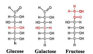
Among other monosaccharides, we can also point out galactose (which is part of lactose or milk sugar) or fructose (present in fruits and honey). Both glucose, galactose and fructose[3] are examples of isomeric monosaccharides because they have the same chemical structure but they differ structurally and chemically due to the different arrangements of their functional groups around their assymetric (or chiral) carbons[4]. Glucose and galactose belong to the group that we saw previously called aldoses and fructose belongs to the ketose group.
Glucose, galactose and fructose molecules

Disaccharides

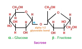
Disaccharides (di-: two) are formed when two monosaccharides undergo a dehydration synthesis. During this process the hydrogen atom of one monosaccharide is combined with the hydroxyl group of other monosaccharide, forming a covalent bond between them and releasing a water molecule. This covalent bond is named glycosidic bond, which is classified in two types: α and β, depending on if the hydroxyl group is located above or below the chiral carbon (also known as asymmetric carbon that we saw previously).
Sucrose molecule: combination of monosaccharides glucose and fructose
Among the most common disaccharides are found lactose, which is the result of the union of galactose and fructose. Milk is a natural source where lactose is found.  Another example of disaccharides is maltose, or malt sugar, which is formed during a dehydration reaction performed by two glucose molecules. But, without a doubt, the most common disaccharide is sacarose, or table sugar, made up of fructose and glucose monomers.


Polysaccharides

Polysaccharides (poly-: many) are long chains of monosaccharides joined by glycosidic bonds. These chains can be branched or unbranched, containing different types of monosaccharides.
Among the main polysaccharides are: cellulose, glucogen, starch and chitin.

Starch
As I mentioned earlier, plants are capable of synthesising glucose from water and CO2. When glucose production exceeds the energy needs of the plant, the surpluses/excesses are stored as starch in different parts of the plant like the root and seeds, providing feed during their germination.
When starch is consumed by animals or humans in their diets, it is first broken down by enzymes (such as salivary amylases) into smaller molecules like glucose, before being absorbed by cells to satisfy their energy needs.

Glucogen
Glucogen, composed of glucose monomers, is how glucose is stored in humans and other vertebrates. It is the equivalent to starch in plants and it is accumulated in muscular cells and the liver. When blood glucose levels decrease, these glucose deposits are demanded and glucogen is broken down into glucose during a process called glycogenolysis.
Glycogenesis and glycogenolysis processes

Cellulose
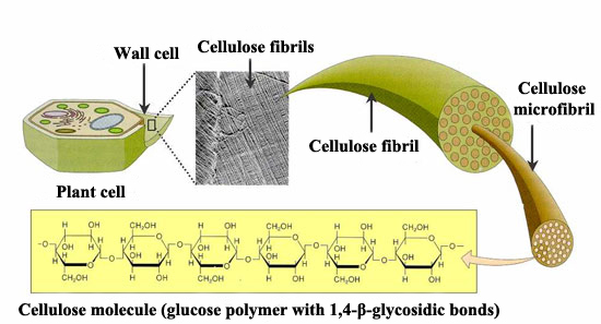
Cellulose, also made up of glucose monomers, is the most abundant polymer. It is the main element of plant cell walls, giving them structural support. In cellulose the basic monomers are tightly packed, giving cellulose its typical fibrous structure which confers stiffness and high tensile strength, i.e. structural support for plants.
Unlike human beings, herbivores have bacteria and protists[5] inside their digestive system which segregate the cellulase enzyme to help digest the cellulose they consume in their grass. By the action of this enzyme, cellulose is broken down into glucose monomers and in this way it can be used as energy source.
Cellulose structure

Chitin
The fourth polysaccharide to point out is chitin, which is the main component in fungi cell walls and the exoskeleton (external skeleton) of arthropods, which protects their internal organs. Chitin is a polysaccharide with a high nitrogen content, constituted by repetitive units of N-acetyl-β-glucosamine, a kind of modified sugar.


BENEFITS OF CARBOHYDRATES

Carbohydrates are composed of both soluble and insoluble elements. Among the insoluble carbohydrates we find fiber, which is mainly cellulose. Cellulose helps to regulate the intestinal transit and the glucose consumption rate. It also binds to the blood cholesterol in the small intestine and in this way the cholesterol is eliminated by being excreted from the organism in faeces. Also, diets high in fibre can help to protect against colorectal cancer.

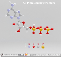
Glucose is provided by the carbohydrates that we consume. Once it is broken down during cell respiration, glucose produces ATP molecules, known as the energy-currency of cells, providing fast energy for any organism.
ATP molecular structure
Consequently, without the consumption of carbohydrates, the availability of instant energy for the body (or any other organism) would be deeply reduced.

For all these reasons, eliminating the ingestion of carbohydrates, like some hypocaloric diets recommend in order to achieve a fast weight loss, is questionably healthy. To accomplish such an aim, it would be more healthy to balance appropriate low GI carbohydrates present in vegetables, fruits and cereals with a balanced level of proteins, vitamins and lipids, a suitable water intake and doing exercise appropriate to our age, gender and physical fitness.


[1] Both are organic compounds characterised by containing the carbonyl functional group (C=O). But aldehydes contain the carbonyl group bonded to at least one hydrogen atom, whereas ketones contain the carbonyl group bonded to two carbon atoms.
[2] Molecule used by all living organism to provide energy in chemical reactions. It is considered as the “energy-currency” of the metabolism.
[3] They all are hexoses: six carbon atoms in their structures.
[4] Carbon atom attached to four different types of atoms or group of atoms.
[5] Eukaryotic (their cell nucleus is delimited by a membrane) organisms, usually unicellular, which due to their own characteristics cannot be included in the rest of the kingdoms (animals, plants and fungi) within this category.

Sources: OpenStax College, Biology. OpenStax College. 30 May 2013.              
              http://medmol.es/glosario/121012glosariomedmol_atp/
              https://kel-tay-lii.wikispaces.com/A.+Intro+to+Phys
              http://chemistry.tutorvista.com/organic-chemistry/oligosaccharides.html
              http://www.entrenasalud.es/glucogeno-y-deporte-un-gran-deposito-de-energia/
              http://www.asturnatura.com/articulos/envoltura-celular/pared-celular.php


Your opinion matters

3.1. Biological macromolecules. Introduction

0 comments
Within this section I will explain the four main types of biological macromolecules: carbohydrates, lipids, proteins and nucleic acids. These macromolecules are the basic constituents of cell structure, that in combining form most of the dry weight of a typical cell. They carry out a wide range of functions in cells. These macromolecules are organic molecules, thus their main component is carbon. Hydrogen (H), oxygen (O) and nitrogen (N) and other minor elements are also part of their structure.
The two fundamental reactions that control the synthesis of biological macromolecules are the following:

    ▣      Dehydration synthesis
The basic blocks of most macromolecules are sub-units known as monomers. These monomers are combined through covalent bonds to form larger molecules (polymers). To do this they release water molecules as byproducts, this mechanism is called dehydration synthesis, which means “to join by losing water”. During these reactions, the hydrogen (H+) of one of the monomers is combined with the hydroxyl group (OH-) of other monomer, releasing a water molecule. At the same time, the monomers share electrons and are joined by covalent bonds.
Different types of monomers or even just a single monomer is combined to form a diverse range of polymers. Thus, for instance, glucose monomers are the components of several different substances such as cellulose, glycogen or starch.
Dehydration reaction

    ▣      Hydrolysis
Hydrolysis (“to separate water”) is a reaction in which water molecules are used to break down the polymers into their basic monomers. In these reactions the polymers are divided in two: one part acquires a hydrogen atom (H+) and the other a hydroxyl group (OH-); both elements (H+ and OH-) occur from the breakdown of a water molecule.
Hydrolysis reaction
Both in hydrolysis and dehydration reactions, enzymes (a specific type of proteins) can act to catalyse (accelerate in these two cases) these reactions.
Through hydrolysis and using particular digestive enzymes, our body breaks down the proteins, lipids and carbohydrates (macromolecules) that we eat. Once these ‘macro’ (i.e. larger) molecules are broken down into smaller molecules, the intestinal cells can absorb the nutrients that they contain. Thus, for instance, lipids are metabolised by lipases, proteins are broken down by the action of hydrochloric acid, peptidase or pepsin and finally carbohydrates are digested by enzymes like lactase, maltase or sucrase.
By breaking down all these molecules into a unit where cells can absorb them as their own ‘food’, these cells obtain the necessary energy to perform all their functions.

Sources: OpenStax College, Biology. OpenStax College. 30 May 2013.               
              http://www.differencebetween.net/science/health/difference-between-hydrolysis-and-dehydration-synthesis/


Your opinion matters

1.2.4. Optimisation of candidates

0 comments
The number of pharmacological compounds that survive the screening phase has to be further reduced and optimised according to key criteria.
To accomplish the largest possible reduction in the number of drug candidates, researchers characterise the way the compounds bind to their targets. This optimisation process is therefore, only applied to the first technique we discussed previously, the target-based approach.
Due to the limited knowledge about the mechanism of action of the chemical compounds in the phenotypic approach, the validation of this optimisation process is quite hard to carry out using this method.
Among the main factors affecting drug-target binding, we find:

      Affinity degree between drug and target.

      How many different targets the drug can bind to.

      How long the chemical compound (drug) can be bound to the target.

These criteria will help to discover the most effective drug dose and detect any undesirable side effects that may occur.
Further, those candidates that present toxicity at therapeutic levels have to be eliminated, as well as those which are highly unstable in solution or solid state, and finally those which are highly expensive to synthesise.
The compounds that meet the necessary requirements are examined by chemists, who produce a certain type of compound called analogs, structurally similar but with improvements in the mentioned criteria.

One of the major tasks during the optimisation process is to determine the correlation between the chemical structure of the compound and its activity (QSAR[1]) and in what way the first factor affects the second one.
Another aspect to evaluate is compound’s drugability, which describes the ability of the drug to reach its target, bind to it and be capable of producing some measurable effect. To find out the drugability of a candidate, scientists use Lipinski’s rule of five, which establishes that an oral medicine fulfils its pharmacological function if:

      It does not contain more than five hydrogen bond donors (total number of nitrogen-hydrogen and oxygen-hydrogen bonds).

      It does not contain more than ten hydrogen bond acceptors (total number of oxygen or nitrogen atoms).

      Its molecular mass is less than 500 uma.

      An octanol-water partition coefficient[2] (log P) less than five.


[1] Quantitative structure-activity relationship is the process whereby the chemical structure of a compound is related to the biological activity of a receptor.
[2] Ratio between the concentration of a certain substance (solute) in two solvents in equilibrium (octanol and water): log P = log ( [solute]octanol / [solute]water ).

Sources: UTAustinX: UT.4.01x Take Your Medicine - The Impact of Drug Development.
              https://es.wikipedia.org/wiki/Relaci%C3%B3n_cuantitativa_estructura_actividad
              https://es.wikipedia.org/wiki/Coeficiente_de_reparto_octanol-agua


Your opinion matters

1.2.3. Screening candidates

0 comments
After the identification and a detailed study of the key target in the development of the disease, there is a second stage based on the screening or selection of the future drugs that will effectively perform on that causal target. This approach dictates the discovery of most marketed drugs presently.
In the selection of chemical compounds, scientists should have an accurate idea about what sorts of candidates are able to bind to the biological receptor in a suitable way, thus minimising the number of compounds that need to be initially screened. This number will be further decreased by the use of specialized software and virtual libraries that contain detailed knowledge about the different pharmacological compounds to be analysed.
Subsequently, the screening carried out in the laboratories uses automated equipment with multiwell plates (carrying the biological receptor), in these plates the performance of the different candidates to study is evaluated in a relatively short period of time.

In principle one would think that the better binding pharmacological compound-target is, the better the expected result should be, but this is not always the case, since knowing the mechanism of disease progression and the role of all its targets is nearly impossible. This is why researchers have developed an alternative approach to this problem, named the phenotypic approach. Using technique a living model[1] of the disease is used and scientists conduct research to see if the candidate has been able to reduce or eliminate the signs of the disease in some way, without knowing the targets and their mechanisms of action.
The main advantage of this method is its higher potential to discover first-class compounds, but not knowing the receptor makes it much harder to modify the chemical compounds. Also, it will be unknown how these likely modifications could affect the binding with the biological target.

Relatively recently, a new type of compounds called biologic compounds have been used, which rather than being produced in a chemical lab, originate from living systems.
Most of them are not going to bind to any targets because they are copies of existing molecules in our organism.
For example, insulin[2] has been produced by genetic engineering by introducing DNA in bacteria colonies. These bacteria grow, multiply and by reading the inserted human genetic code produce the insulin.
Some of these biological compounds can also perform over receptors, because tailor made proteins and antibodies can be designed to bind or interfere with those elements that cause diseases in cells.


[1] These living models are cells, cellular tissues or even mice developed to model human diseases.
[2] Hormone made up of 51 aminoacids, produced and secreted by pancreatic beta cells. It is in charge of regulating glucose levels in blood.

Source: UTAustinX: UT.4.01x Take Your Medicine - The Impact of Drug Development.


Your opinion matters

1.2.2. Identifying targets

0 comments
The development of a new drug is initiated with the identification and the selection of biological receptors (targets) on which that pharmacological compound will perform on. These targets are ordinarily proteins where the compound binds to produce changes in their functions.
Proteins are the workers which perform most of functions in human cells, consequently it makes sense that these are the places where drugs bind to transform their activities, modifying this activity will help to eliminate or, at least, reduce diseases in infected cells.
Genes can be a target for drugs as well, which are in charge of providing instructions about what proteins must be created and what tasks they will fulfil within the cell. These proteins and genes are not always human: it is estimated that around 15-20% of current medicines act on proteins and genes of the pathogen that has invaded our body and caused diseases.

The problem related to many diseases is the fact that hundreds of genes and proteins can be involved in their progression, that is the reason why the selection of the key receptor by the researchers is a fundamental factor.
To carry out this task it is essential to statistically analyse a huge amount of data to be able to choose the most suitable biological target for the chemical compound.

On some occasions, just the binding between a drug and a target is enough to eliminate a disease. For instance, amoxicillin antibiotic binds certain proteins on cell walls of the infectious bacteria, leading to the breakdown of these walls and therefore, the death of these bacteria. However, it is not always so easy, since those bacteria that have developed antibiotic resistance have been able to modify the receptors where drugs perform, losing, in this way, their therapeutic action.
Often, other diseases are really hard to overcome because they are caused by a combination of genetic, environmental factors and lifestyle factors: Alzheimers being one example.
Lastly, at other times, it may be more appropriate to adopt a “multi-target” approach where several drugs are administered to a single receptor, where a single drug performs on multiple targets, or a combination of both techniques.

Source: UTAustinX: UT.4.01x Take Your Medicine - The Impact of Drug Development.


Your opinion matters

1.2.1. Introduction to drug discovery. Prediscovery of targets

0 comments
In past, research into new drugs was accomplished in a very different way to today. Many of these drugs were simple homemade remedies, which were passed on from generation to generation and most of them discovered by accident. This is the case for one of the most famous antibiotics ever: penicillin. 
In 1928, Alexander Fleming observed that a staphylococcus bacteria culture that he was studying was
contaminated by a certain fungus (penicillium) and that the area around these fungi was free from bacteria. It was later on, Howard Floyd who realised the tremendous potential of penicillin for wounded soldiers during the II World War.
Although these kinds of discoveries still occur today, they are not as usual as they used to be and tend to have less impact than penicillin had at the time. Currently a much more methodical process is followed which involves industry, universities, government agencies and even philanthropic organisations. The development of drugs begins with the discovery of new drugs, which starts with approximately 10,000 potential candidates of which 9,999 will be ruled out until the best of these compounds is selected. The chosen candidate will have a suitable equilibrium between efficacy and safety. The discovery phase can also be broken down in four stages:

      The pre-phase of research about the disease on which the future drug will act.

      Identifying a target disease where onto the drug can bind (known as biological or therapeutic target).

      Screening of the chemical compounds that perform on these targets.

      Optimization of these compounds until they produce the lead candidate.
Drug development stages


PREDISCOVERY OF TARGETS

As you could see from my previous post in this section, drug development is a really long process with high costs both in terms of human effort and financial investment. You can imagine then that before beginning this process we need to answer a set of questions to give us a clear idea about the next steps to take:

      What are the unmet medical needs today, what drug can be developed that allows us to establish a solution to a certain disease whose treatment or cure does not exist or is deficient currently.

      To have a broad knowledge about the illness which the drug will work on. This knowledge includes the causes of the disease, which is not always trivial.

      What proteins and genes are altered by the disease and how that affects the way in which these genes encode proteins.

      How cells and tissues are also modified.

      Finally, how the disease affects the whole patient.

Hence, this is a study from the smallest target where the disease can act (proteins and genes) to the largest (the patient).

Sources:  UTAustinX: UT.4.01x Take Your Medicine - The Impact of Drug Development.
               UC San Diego: Drug Discovery, Development & Commercialization.


Your opinion matters

1.3. Top-down and bottom-up approach

0 comments
Current manufacturing technology is based on the approach known as “top-down” which consists of, in a similar way to a sculptor does, starting from a large block and shaping and chiseling it, to obtain, progressively, a smaller object with a desired shape.
In the nano approach, we progress in the opposite way, from the small structure, building up to a bigger one. This method is called the “bottom-up” approach, in which like lego (with a large number of pieces with different shapes, colours and size), we are going to begin with basic elements such as atoms, nanoparticles, nucleic acids or proteins to assemble molecules or even to build diverse sensors and devices.
To enable manufacturing using nanotechnology, special tools which enabled the visualization as well as the manipulation of objects at nanoscale are required. The scanning probe microscopy (SPM), for instance, accomplishes this task by allowing us not only to observe but also to move atoms on a surface.

The progress of nanoscience and nanotechnology and the use of this highly advanced instrumentation created one of the most significant features of this field: it is multidisciplinary. By reducing the scale, atoms and molecules become the basic “bricks” which physicist, chemists, biologists and engineers work with, using a common language. One example of this multidisciplinarity is the design and manufacturing of a biosensor where a biologist must have knowledge of quantum physics and a physicist about biology in order to design their end product successfully.

Assembly on a molecular or particle basis to develop all the technology we demand may seem unrealistic, nevertheless this is what the Earth has been doing for the last 4 billion years, since starting from simple molecules it formed really complex structures by linking and auto-assembling substances. Any living organism is, undoubtedly, a clear example of bottom-up building; starting from certain organic molecules and a genetic sequence it has been possible to create very complex structures functionally and structurally. As a consequence, nanotechnology can learn from these processes to imitate and adapt them, even, to other kinds of problems very different to biology.

The development of nanoscience and nanotechnology, according to experts, will occur in three stages. The firt one between the years 2000 and 2020, where industries will mainly keep using conventional production techniques (top-down). The second stage between 2010 and 2030, where bottom-up methodology will begin to spread, and ultimately become the leading manufacturing scheme for the rest of the XXI century. But this fact does not mean that our current procedures will totally disappear, because the use of one system or the other will be dependent on many factors like raw materials, labour, environmental and social costs and, of course, economic profitability.
Nanotechnology is already a major industry, worth 50 billion dollars globally market in 2006, with expected growth to a trillion dollars in 2015. Therefore, this market will benefit those companies whose countries are investing in this field long term.


Sources: Fundación española para la ciencia y la tecnología (FECYT). Nanociencia y nanotecnología. Entre la ciencia ficción del presente 
              y la tecnología del futuro, 2009.
              http://researcher.watson.ibm.com/researcher/view_group_subpage.php?id=4252
              http://www3.nd.edu/~kamatlab/facilities_physchar.html


Your opinion matters

1.2. Nanostructures

0 comments
Nanostructures can be defined as those objects where, at least one of their dimensions is located at nanoscale (1-100 nm). Among the most commonly used structures in nanomedicine field we find:

      Micelles: structures with spherical or globular shape. They are made of molecules that have a polar or hydrophilic head (with strong affinity for water) and a non-polar or hydrophobic tail (repels water). The heads are placed in the outer region of the micelle forming a layer in contact with the liquid environment surrounding it; the tail, instead, is located in the interior forming the nucleus of this nanostructure.
Their typical size is c50 nm and they are used to carry and deliver water-insoluble drugs, which are confined inside the hydrophobic core of the micelle, in such a way that they are protected from the exterior aqueous environment.
One of their most interesting features is that they can circulate through the bloodstream for longer than other sorts of particles because they can avoid macrophage[1] action.

      Liposomes: closed vesicles made up of lipid bilayers (two lipid layers faced by their hydrophobic tails). According to the number of these bilayers the liposomes may be classified as unilamellar or multilamellar.
Unilamellar liposomes are characterised by an aqueous core to carry water soluble drugs. Multilamellar liposomes do the same thing with liposoluble drugs. When they are intravenously administrated, liposomes are cleared quickly by the reticulendothelial system (RES)[2], in addition, hydrophobic, electrostatic  and Van der Waals forces may disintegrate them. To avoid this, these nanostructures are coated with inert polymers, like polyethylene glycol (PEG) which enables their circulation through the body without being excreted.
Some liposomes are designed to be degraded only where necessary, for instance, in low pH areas (tumour regions with hypoxia[3]), others are functionalized with antibodies or ligands (molecules that bind specific celullar receptor) onto the nanoparticle surface so that they can bind and perform on these receptors. A third technique is based on the development of thermo-labile liposomes, which are guided to target tumor tissue by hyperthermia.

      Dendrimers: three-dimensional systems with treelike structures. These nanostructures consist of a central core molecule with lots of branches. Their shapes and sizes are controlled in a very accurate way by polimerization from the central core or by synthesis from the periphery to the core molecule.
Dendrimers are excellent candidates for carrying drugs, due to the fact that they offer a high stability and their functionalization (attachment of functional groups on the surface of the nanoparticle) by physical or chemical interactions.
They can deliver a wide range of molecules, both hydrophilic and hydrophobic ones, anticancer agents, drugs or contrast agents for diagnostic imaging.
Types of nanostructures

      Nanospheres: spherical structures made of matrix systems in which the drug is distributed by encapsulation, entrapment or attachment. The nanosphere surface is modified by the addition of biological material (antibodies or ligands) or polymers so that they can reach their targets in cells.

      Nanocapsules: vesicle systems where the drug is confined in a cavity or nuclear core, surrounded by a polymeric membrane where ligands and antibodies can be attached. The core material may be solid, liquid or even gas, always in an aqueous or oily environment.

      Carbon nanotubes: mono or multilayer cylindrical structures constituted by graphite or another carbon material. They deliver their loads in a specific way by functionalizing their surfaces with nucleic acids, proteins or bioactive peptides[4], which allows them to become particles with a very low toxicity. They are good candidates for carrying and delivering drugs because they are not immunogenic (they do not produce an immune response).

      Polimeric nanoparticles: are one of the most adequate and suitable material used in nanomedicine, being largely biocompatible and biodegradable. Among their advantages are: the potential to modify their surfaces by chemical transformations, the encapsulation of the load to transport, carrying a broad variety of therapeutic agents and lastly an outstanding pharmacokinetic control[5] of these agents.
Their polymeric coating reduces immnunogenicity and limits their phagocytosis by the reticuloendothelial system (RES), increasing, in this way, the blood levels of the carried drugs in organs like the brain, intestines and kidneys. They are normally designed in such a way that are sensitive to the environment, delivering their carried drugs by responding to both physical stimuli (temperature, solvents, light) and chemical stimuli (reactants, pH, ions in solution or chemical recognition).

      Inorganic nanoparticles: usually composed by (SiO2) or alumina (Al2O3).  However, their cores are not just limited to these two materials, they can be made of any kind of metal, oxides and metal sulfides, which leads to a myriad of nanoparticles with a large range of shapes, sizes and porosities.
They are normally produced to avoid RES by modifying their size and superficial composition. They are porous with a physical coating that protects the carried load from a likely degradation or denaturation.


[1] Large size cells of the immune system located in different organs.
[2] System composed by a group of cells whose function is to capture inert particles in the body.
[3] Condition in which blood, cells or tissues is deprived of sufficient oxygen supply.
[4] Molecules formed by the union of several amino acids.
[5] Control of the processes that a drug is undergo within the organism.

Sources: Barbara Haley, Eugene Frenkel, Nanoparticles for drug delivery. Elsevier, 2008.
             Jose Manuel González, Marta López, Gema Ruiz. Informe de vigilancia tecnológica, nanomedicna 2006
             Amir H. Faraji, Peter Wipf. Nanoparticles in cellular drug delivery. Elsevier, 2009


Your opinion matters

2.5. The carbon atom

6 comments
Cells are made up of a wide range of complex molecules, called biological macromolecules, like carbohydrates, lipids, proteins and nucleic acids (which I will deal with in my next posts). In this post we will discuss carbon, the basic element of all these macromolecules.
Carbon (C), atomic number 6 (6 protons and 6 electrons), has an incomplete outermost shell with four electrons in it. Thus, with these four unpaired electrons, it can form up to four covalent bonds with other atoms satisfying, in this way,  the octet rule. This property allows carbon to become a really versatile chemical element, ideal as a structural component or as the backbone in macromolecules.


Hydrocarbons

Hydrocarbons are organic molecules[1] completely made up of carbon and hydrogen. The covalent bonds that are formed between the atoms of these molecules release a great amount of energy when they are burnt (oxidized). This is the reason why hydrocarbons are used as fuels in our daily life. Some examples include butane and propane gases.
Each covalent bond between carbon atoms can be simple, double or triple, which influences the geometry and global shape of the molecule, which in turn, influences the properties and functions of hydrocarbons in a basic way. Thus, we find that carbon atoms with simple bonds constitute tetrahedral shapes, which allows the rotation of the molecule along the axis of the bond. However, when double and triple bonds appear, the molecule configuration is planar and linear respectively.
Hydrocarbons are divided, mainly, into two categories:

      Aliphatic hydrocarbons (hydrocarbon chains): they are made up of successive bonds between carbon atoms which can be branched or unbranched.

      Aromatic hydrocarbons (hydrocarbon rings): they are formed by closed rings of five or six carbon atoms. These kinds of structures occur, at times, in double bond hydrocarbons, like cyclohexane or benzene. Benzene has vital importance in some amino acids, like the cholesterol molecule or its derivatives (testosterone and estrogen hormones).
Nevertheless, this division is not definitive because some hydrocarbons present their structure with both aliphatic an aromatic fragments, an example is the beta carotene molecule, which is a powerful antioxidant and precursor of A vitamin, present in fruits, vegetables and grains.


Isomers

Isomers are molecules that share the same chemical formula but with a different structure or placement of their atoms and/or chemical bonds. We can distinguish two types:

      Structural isomers: they differ on account of the situation of their covalent bonds. Thus, butane and isobutane are constituted by 4 carbon atoms and 10 hydrogen atoms each (C4H10), but the different placement of the atoms within the molecule leads to different chemical properties. Whereas the first one (butane) is used as a fuel, the second one is more suitable to use as a refrigerant and a propellant in sprays.

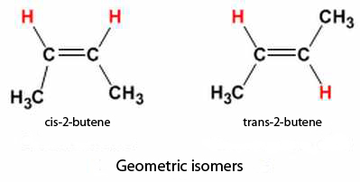
Geometric isomers: they are differentiated by how the atoms are configured around double carbon bonds C=C. Thus, we would talk about cis configuration, when we find the same groups of atoms at the same side of the double bond, and trans configuration, when they are arranged on opposite sides. Trans configuration generates an approximate linear molecular structure, whereas the cis configuration originates a bend (change in direction) of the backbone.
Cis and trans configuration of isobutene

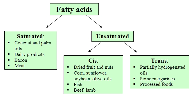
Triglycerides[2] (fats and oils) are classified according to the fatty acids[3] they contain. Thus, those fatty acids with at least one double bond between carbon atoms are unsaturated fats. When some of these bonds appears with cis configuration, the triglycerides molecules cannot be grouped or packed, so they are liquid at room temperature constituting those substances known as oils.
However, if the double bond  presents trans configuration, the molecules are able to pack tightly at room temperature forming solid fats, popularly known as trans fats, such as partially hydrogenated oils, processed foods and some margarines. In the human diet, these fats are associated with an increase in cardiovascular diseases, therefore they have been significantly reduced and/or eliminated in lots of food products in food sector.
In contrast, those triglycerides that lack double bonds between carbon atoms are named saturated fats, meaning that they contain all the hydrogen atoms available. These fats usually solidify at room temperature and are found in food of animal origin, milk and its derivates, and even in some oils of plant origin, such as coconut and palm oils.
Classification of fatty acids

Enantiomers

Enantiomers are molecules with the same bonds and identical chemical structure but with different arrangement of their atoms, such that they are mirror images of each other. An example is some D and L- forms of amino acids which have very different functions, the D-form of amino acids composes the cell walls in some bacteria, and the L-form of amino acid makes proteins.
L- and D- amino 
acids


Functional groups

Functional groups are groups of atoms in molecules, which give them some specific chemical properties. In this way, each one of the four groups of biological macromolecules (carbohydrates, lipids, proteins and nucleic acids) has its own set of functional groups which provide them chemical properties and very particular functions in living organisms.
These groups are attached to the carbon backbone at several points in macromolecules along their linear chain and/or ring structure.
Functional groups are usually classified on the basis of their charge or polarity, in hydrophobic (uncharged molecules that do not interact well with polar molecules like water) and hydrophilic (polar ions or molecules that do interact well with other polar molecules).


[1] Those molecules that contain any form of carbon (solid, liquid or gas) which are vital for life.
[2] Compound made of a glycerol molecule as a backbone with three fatty acids attached.
[3] Long aliphatic chains usually containing an even number of carbon atoms (16 - 22). Their structure is mainly hydrophobic (repels water) with a carboxylic group as a functional group with acidic character.

Sources: OpenStax College, Biology. OpenStax College. 30 May 2013.              
              http://perdergrasa.bligoo.es/clasificacion-de-trigliceridos
              http://recursos.cnice.mec.es/biosfera/alumno/2bachillerato/biomol/contenidos9.htm
              http://biomodel.uah.es/model2/lip/acgr-salud.htm


6 comments

Your opinion matters

1.3. Basic and applied science

0 comments
For a long time, and even now, the value of science has been a topic of debate. This debate has broadly divided science into two categories according to its goals, but not necessarily 'value':

      Basic science or pure science: the search for knowledge regardless of its possible applications in the short run. Its immediate goal is knowledge just for knowledge's sake, no matter whether there is a practical application or not.

      Applied science or technology: whose objective, on the contrary, is to enable solutions to everyday problems, which, as a general rule, are defined by researchers.


Basic science. What value?

There are some people who define applied sciences as “useful” and basic sciences, however, as “useless” and therefore deserving of less attention. However, if we have a look at the history of science, basic knowledge has enabled, subsequently, the development of remarkable applied science. I will list examples below:

Applied scientific discoveries at the famous CERN (European Nuclear Research Centre) initially can be seen as purely theoretical, but contrary to what most people think, these discoveries have day to day applications like the Internet. For example, discoveries at CERN allowed the development of the world's first web site. Also within the computing world, CERN has made feasible the development of GRID, which is a distributed computing system capable of managing the 15 million GB of data that the CERN generates each year. This GRID system allows the distribution of this amount of information and its access by researchers all over the world.

In the medical field, the development of particle accelerators like LHC (Large Hadron Collider) at CERN has provided the emergence of techniques for the treatment of certain diseases like cancer.
One example of these practical applications is hadrontherapy where the tumour is bombed by protons, which enable both more accurate cancer therapy as well as one with reduced patient side effects. Great progress has also been achieved in noninvasive diagnostic imaging techniques like PET (positron emission tomography).

The environment benefits from the advances accomplished at CERN as well, since the complex electronic systems used for detecting particles in accelerators can be applied in environments in which there is the risk of a radiation leak like nuclear power plants in order to enable early detection.
The former accelerator to LHC, LEP, was built using plastic that did not contain sulphur or halogen compounds, which in case of fire, did not produce extremely toxic fumes. These new non-toxic plastics have now been adopted extensively by industry.

Whilst it is arguable that disproportionate amounts of money are spent in order to enable these discoveries, nonetheless I believe that a large number of solutions could not be found without a broad theoretical knowledge produced by basic science.

Source: OpenStax College, Biology. OpenStax College. 30 May 2013.
            http://elgrancolisionadordehadroneshoy.blogspot.com.es/2014/06/aplicaciones-practicas-posibles-riesgos.html
            http://cern123.galeon.com/Beneficios.html             


Your opinion matters

1.1. Nanotechnology. Introduction

0 comments
It is very likely that the term nanomedicine has caught your eye and maybe this is the first time you have heard of it. As nanomedicine can be defined as the application of nanotechnology in the medical field, I will begin this section with a brief view about what nanotechnology is. I will show you the unique features of technologies and materials at nanoscale and a classification of the most common types of structures used in nanomedicine.

But first of all, what is a nanometre?, the prefix “nano” comes from the Greek νάνος, which means “dwarf”. Nowadays, it indicates the billionth part or, in other words, it is a factor of 10-9, therefore, a nanometre (nm) is one billionth of a meter. This way, we can conclude that nanoscience/nanotechnology is the science/engineering that studies and operates with matter in a size from 1 to 100 nm.

The idea and concept of nanotechnology arose from a Richard Feynman’s conference at UCLA University in 1967, where this theoretical physicist introduced, for the first time, the possibility of manipulating atoms and molecules.
But nanotechnology age really began in 1981 when the scanning tunneling microscope (STM) was developed with which we are capable of observing atoms.
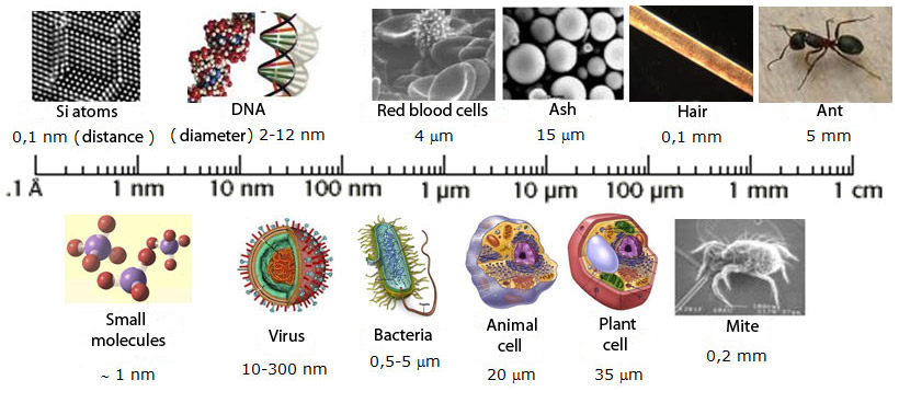
The smallest objects we can observe with our naked eye have a size around a millimetre (the thousandth part of a meter), e.g.: the edge of a coin or a grain of sand, below this magnitude we find it difficult to distinguish objects.
If we divide a millimetre into a thousand parts we are in the micrometer scale, which is the domain of bacteria (5-20 μm) or blood cells (red blood cells: 6-10 μm). Therefore, to observe them we need the help of an optic microscope.
If we keep decreasing the scale and we cut up a micrometer into a thousand parts, we achieve our goal: the nanometre, as I said previously the billionth part of a meter. At this scale we find viruses (20-250 nm) and the DNA molecule (around 2 nm wide).
However, to be able to observe an atom we should still decrease one order of magnitude our scale, as atoms have magnitudes from 0.1 to 0.3 nm.
The scale of things
To get a more accurate idea about the real size of a nanometre, here are a couple of objects from our daily life measured in nanometres: a human hair is about 50,000-100,000 nm in diameter and a paper sheet is around 100,000 nm thick. Now, let’s do it the other way round, imagine you are shrunk until 10 nm, at that scale a human hair is like the island of Manhattan, a red blood cell like a football stadium, a polio virus like a basketball hoop and a hydrogen atom like a ping-pong ball…...¿surprising?

Right now, you may be wondering what the point is of using such tiny scales and you can find the answer, for instance, in your mobile phone. Miniaturisation has transform the huge, old mobile phones into small computers (with GPS, Internet connection, digital camera…) that you can carry in your pocket.
Nanotechnology has also enabled several breakthroughs like remote medical diagnostic devices, holograms, flexible and 3D screens, or seamless voice control devices.
Therefore, miniaturisation has allowed to locate millions of electronic devices in an area of just a few millimetres.


Surface area to volume ratio

One of the most important properties at nanoscale is the surface-volume ratio. This ratio is an essential parameter in miniaturization and nanotechnology, which states that this ratio increases as we decrease the dimensions of an object and vice versa.
As a material size diminishes, most of its atoms are located on the surface. Let’s consider a 10 nm silicon[1] cube, if we make some calculations we find that it contains around 50,000 atoms in all, of which 680 are located on each face of the cube. So, the overall number of atoms on the surface, multiplying by six, comes to 4,080. Now, if we divide this number by the total amount (50,000 atoms) we obtain that around 8% of the atoms are on the cube surface.
Let’s carry out the same calculations with a 10 cm2 and 1 μm thick cube, we get, in this case, that only 0.03% are found on the surface.
Therefore, from these calculations we can conclude that nanomaterials have a greater surface area per unit volume than larger materials. This ratio leads us to a really interesting property in materials at nanoscale: they are much more reactive from a chemical point of view, so they can catalyse reactions more easily, why? because atoms and molecules on surfaces do not have full allocation of covalent bonds, consequently, they are energetically unstable what makes them be more reactive than the non-nanoscale materials.

Because their specific physicochemical properties, nanomaterials have innumerable applications since they can participate in biological processes interacting with biological macromolecules (such as carbohydrates, nucleic acids, lipids and proteins). Also with ions, minerals, water in desalination treatments or even in drug delivery on which particularly I will focus on later.
Hence, there is a paradigm shift in nanotechnology: what is important about materials is not really what they are made of, but how small they are.


[1] Compound of oxygen and silicon ordered in a three-dimensional structure forming quartz and its types.

Sources: Introduction to Nanotechnology, Prof. Hossam Haick, Israel Institute of Technology.
              Rice University. Nanotechnology: The basics.
              http://www.quimicaviva.qb.fcen.uba.ar/v11n3/castro.html


Your opinion matters

1.1. Drug development. Overview

0 comments
First of all, to begin this section I would like to describe a brief overview about the different phases in drug development that we will study in more detail as we move forward.
Drugs are one of the substances that we are more familiar with in our daily life and we consume them more or less often depending on our health condition. However, very few people know their manufacturing process and the huge effort needed so that they can reach the market and our hands.
All around the world there are thousands of research teams analysing diseases and trying to find out their origins and causes. Once causes have been found, biopharmaceutical companies start to investigate how they can act to stop them or even revert their advance. To accomplish this goal tens of thousands of compounds are tested, whose origin can be:

      Natural: from substances that already exist in nature like morphin (plant origin), penicillin (microbial origin) or trabectedin (anti-tumor drug of marine origin).

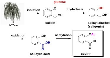
      Semisynthetic: compounds obtained in the lab like acetylsalicylic acid, synthesised from willow bark.
Aspirin manufacturing process

      Synthetic: compounds like ibuprofen with a complete chemical synthesis.

      Biotechnological: these drugs are manufactured with biotechnological techniques where the genetic material of bacteria is modified to produce substances with pharmaceutical interest such as human hormones (insulin) or antibodies.

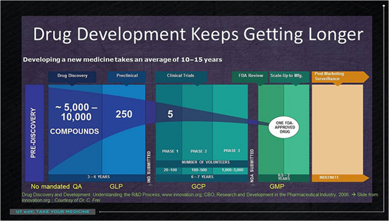
Through a hard and long screening process, that can take over six years, just a few drug candidates are selected (around 10 out of 10,000 initial substances).
All the information about their toxicity and activity in cells and experimental animals is gathered (this phase is called “preclinical phase” where good laboratory practices are followed: GLP). With this information the company requests authorization a drug regulatory agency to initiate the clinical trials. If this authorization is given, the drug candidates are subject to a clinical trial (according to the good clinical practice guidelines: GCP) divided into three stages. Due to the emphasis on safety and effectiveness not all candidates cannot move beyond this long process successfully. These three stages are the following:

      In phase 1 the drug is tested on human beings for the first time. Small trials, which are focused on safety and how the drug is distributed in the body, are performed on healthy volunteers.

      In phase 2 the trial is conducted at a larger scale on volunteers actually suffering from the disease of interest. In this stage the main concern is drug effectiveness and the right doses.

      In phase 3 the trials are carried out on a large number of people in order to enhance the acquired knowledge in the former phase with a larger and broader range of people, involving a lot more money as well.
Drug development phases
If the clinical trial succeeds, which takes around 6-7 years, the biopharmaceutical company processes a new application that will be reviewed thoroughly for a long period of time.
Once the evaluation is over, only safe and effective drugs will be authorised for public use, but despite this approval the regulatory agency will keep monitoring the drug in the market.
Not only during the preclinical and clinical stages strict regulations are followed, but also during the manufacturing process. These rules and regulations are known as GMP guidelines (good manufacturing practices).
Although the final result of this process can be as simple as taking a pill with some water twice a day, it requires a total period of time from 12 to 15 years, an investment between $1-$2 billion and thousands of people who are involved to develop a single drug.

Source: UTAustinX: UT.4.01x Take Your Medicine - The Impact of Drug Development.


Your opinion matters