• Biology

    Biology

    The atom, biological macromolecules (carbohydrates, lípids, proteins, nucleic acids), cell biology, cancer

  • Drugs

    Drugs

    Drug development, pharmacodynamics, pharmacokinetics, toxicology

  • Nanomedicine

    Nanomedicine

    Introduction to nanotechnology, diagnostic devices, drug delivery, regenerative medicine

  • Miscellaneous

    Miscellaneous

    A more in-depth explanation of topics previously mentioned in the other sections

1.4.1. Clinical research: Overview

0 comments
Those compounds that have passed the preclinical phase are tested in human beings (clinical research).
This kind of research has significant associated issues of a scientific, ethical, social and economic nature.

In all developed societies it is mandatory to prove the efficacy and safety of drugs before commercialising them, a task that is fulfilled by clinical trials.
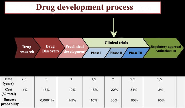
Phases of drug development
Clinical trials are defined as research studies in human beings to determine or confirm clinical, pharmacological, pharmacodynamic effects and/or detect adverse reactions and/or study the absorption, distribution, metabolism and excretion of a drug in order to establish its safety and efficacy.

Clinical tests are based on documentation, both national and international regulation and a series of ethical codes, declarations and international conventions.

Over the last fifty years hundreds of thousands of compounds have been produced, but only 10% of them have passed clinical evaluation, and even within this group some of them were withdrawn after being put on the market due to safety and efficacy reevaluations.
Stages of new drug development
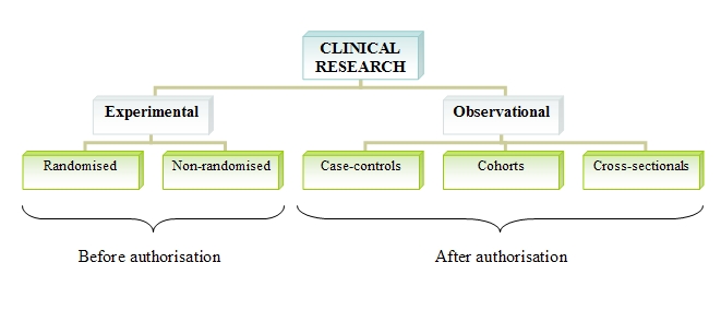
The methodology of clinical research can be carried out via one of the following procedures:
      Experimental: In experimental tests, researchers design the study, select the patients, establish the treatments and variables to study.
      Observational: Researchers are mere observers, without altering or intervening the habitual clinical practice.

Within the experimental tests, we find:
      Randomised trials. In these trials, patients are allocated to one treatment or another (often a placebo) in a random way. This is the procedure demanded by regulatory agencies to avoid bias in data interpretation before giving the authorisation to market the drug.
      Non-randomised trials. Here, patients are not allocated randomly.

Among the observational studies to develop the complete efficacy and safety profile of a drug after its commercialisation, we have:
      Case-control studies. Participants are chosen according to they have or not a certain disease and it is investigated if they were exposed or not to a factor of interest. The exposed group and the control group are compared.
      Cohort studies. The frequency of a sickness is compared between two populations, one exposed to a risk factor and the other population are not.
      Cross-sectional studies. They examine the relationship between an illness and a set of variables in a certain population at a given point in time.
Types of clinical trials
Clinical trials chronologically go through three phases until achieving marketing authorisation for the drug. Once the drug is commercialised, it begins its phase IV, which lasts the whole lifespan of the drug. In Phase IV the drug can be removed from the market if undesired effects, not detected in the three previous stages, are identified.
These new side effects can be discovered when the drug is distributed among thousands of patients.

The measured efficacy in these real conditions is called effectiveness to distinguish it from the efficacy measured prior to drug distribution on the market.

Sources: CEU Universidad San Pablo: Farmacología Básica, 2013.
              http://cmidd.northwestern.edu/about/drug-discovery-and-development-process/
              http://indacea.org/desarrollo-de-medicamentos-1/


Read more

3.5. Nucleic acids

2 comments
DNA and RNA are the most important biological macromolecules for the continuity of life.
DNA molecules contain the genetic material of all living organisms, from bacteria to multicellular mammals.
In prokaryotic cells DNA is not enclosed inside a membranous envelope, but is in eukaryotic cells where it is located inside the nucleus and organelles.

In eukaryotic cells, DNA along with some proteins, called histones, form chromatin (the material from which eukaryotic chromosomes are made), which can contain tens of thousands of genes.
DNA is in charge of controlling the activity of cells by activating and deactivating genes.

The other kind of nucleic acid, RNA, primarily conducts protein synthesis and its regulation through the action of three different sorts of RNA (rRNA or ribosomal RNA, tRNA or transfer RNA and miRNA or microRNA). The fourth type of RNA (mRNA or messenger RNA) acts as communication intermediary between DNA and the rest of the cell.
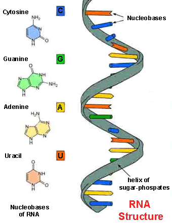
DNA structureRNA structure
Nucleotides are nucleic acid monomers, which are combined to generate their polynucleotides (DNA and RNA). Each nucleotide is composed of three components: one nitrogenous base attached to a pentose sugar molecule, which, in turn, is attached to a phosphate group.

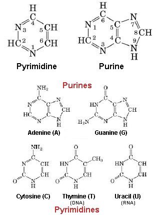
Nitrogenous bases are organic molecules made up of carbon (C) and nitrogen (N) that are divided into five kinds: adenine (A) and guanine (G) are known as purines, they have two fused rings of C-N. Cytosine (C), thymine (T) and uracil (U) are known as pyrimidines, they only have a fused ring of C-N.
Both purines and pyrimidines have different functional groups attached to these rings.

The DNA molecule comprises A, T, G and C, while the RNA molecule comprises A, U, G and C, therefore, tymine is substituted by uracil.
Types of nitrogenous bases
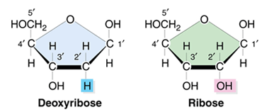
Another difference between DNA and RNA is that DNA sugar molecule is a deoxyribose (they have a hydrogen atom attached to the second carbon atom of the molecule) whereas RNA sugar molecule is a ribose (they have a hydroxyl group attached to that second carbon atom instead).
Deoxyribose and ribose molecules
Carbon atoms of the sugar molecule are numbered as 1´, 2´, 3´, 4´ and 5´. The phosphate residue joins the hydroxyl group of the 5´carbon and the 3´ carbon of the sugar molecule of the next nucleotide forming a phosphodiester linkage between them.


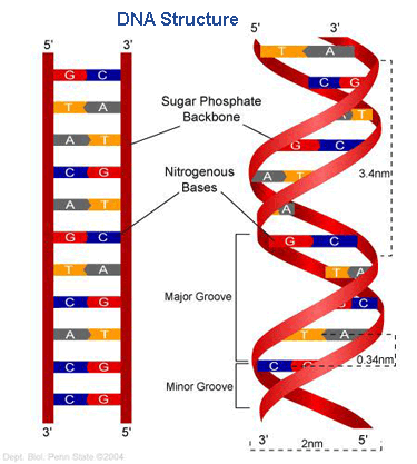
DNA double-helix structure

In the DNA double-helix structure, the sugar molecule and phosphate group are located outside this structure, whereas the nitrogenous bases lie in the interior like the rungs of a ladder linked by hydrogen bonds, in such a way that A can only pair with T and G can only pair with C. This rule is known as the rule of complementary base pairing.
Each of these pairs is separated from the next one by 0.34 nm; both strands of the double-helix run in opposite directions in such a way that the 5' carbon of one strand faces the 3' carbon of the complementary strand.

During DNA replication, each of these strands are separated and they are used to synthesise a new complementary strand, in such a way that each daughter DNA double helix contains a new synthesised strand and one parental DNA strand.


RNA

Normally, RNA comprises a single ribonucleotide strand held together by phosphodiester bonds. These ribonucleotides contain, in turn, a phosphate group, a ribose molecule (pentose sugar) and one of the four nitrogenous bases (A, U, C and G).

The DNA that controls the activity of the cell uses the mRNA (messenger RNA) to carry its message
Thus, if a cell requires a certain protein, the gene for this product is activated and the nucleus synthesises the mRNA. This mRNA has a complementary sequence to the coding sequence of the DNA has been copied from.
The mRNA crosses the nuclear membrane, gets to the cytoplasm and there it interacts with the ribosomes and other cellular machinery.
To produce the required protein, the mRNA is read in groups of three bases called codons. Each codon codifies a single amino acid.

The second kind of RNA, rRNA, is responsible for the suitable alignment between the mRNA and ribosomes. It also has an enzymatic activity and catalyses the formation of peptide bonds between two aligned amino acids.

The tRNA is in charge of transporting the correct amino acid to the site where the protein synthesis takes place. The base pairing between the tRNA and the mRNA allows for the appropriate amino acid to be inserted in the polypeptide chain.

Lastly, the major function of the microRNA (the smallest of all types of RNA) is the regulation of gene expression by interfering with the expression of certain messages of the mRNA.
Translation process
Hence, in an organism the information goes from DNA to RNA and from this one to proteins. During the first stage (transcription), the DNA dictates the structure of mRNA and during the second (translation), RNA rules the structure of a specific protein.
This process is known as the central dogma of molecular biology, which is valid for all living organisms except for some viral infections.
Protein synthesis process

Sources: OpenStax College, Biology. OpenStax College. 30 May 2013.
              https://www.windowssearch-exp.com/images/search?q=Antiparallel+DNA+Replication&view=detailv2&&id=CE5B36CC30
              3DB6E9F7C902454DF516328A0882B8&selectedIndex=1&ccid=uEIdkd5u&simid=608029733518446573&thid=OIP.Mb84

              21d91de6e5ee9db88b338c5b77e9aH0&ajaxhist=0
              http://www.scienceprofonline.com/genetics/ribonucleic-acid-rna-structure-and-function.html
              http://guia.bio.br/tag/purinas/
              http://www.discoveryandinnovation.com/BIOL202/notes/lecture12.html
              http://www.ib.bioninja.com.au/standard-level/topic-3-chemicals-of-life/35-transcription-and-transl.html
              http://thelessonlocker.com/kvhs/biology/biology.html


Read more

2.2. Atomic force microscope (AFM)

0 comments
In the post titled “Top-down and bottom-up approach” I mentioned that, in order to examine and handle objects at the nanoscale it is necessary to develop devices that enable observation of samples at the atomic scale. These instruments are called scanning probe microscopes (SPMs), from which 3D images are obtained at a very high resolution without damaging the analysed sample (an improvement on electron microscopes (TEMs)).
The scanning probe microscopes are divided, basically, into two types: scanning tunneling microscope (STMs), which we studied in the previous entry within this section, and the atomic force microscopes (AFMs), which we address below.
Atomic force microscope (AFM)
Atomic force microscopes are based on the following operating principle:
The sample topography is scanned by a microcantilever or strip around a few hundreds of microns (micrometres) long, built in silicon or silicon nitride, whose end has an extremely sharp tip and a curvature radius on the order of nanometers.

When this tip approximates the sample surface, the forces between them produce the cantilever deflection according to Hooke’s law[1].
Depending on the type of tip-sample interaction (whether there is contact or not), the forces measured by AFMs can be: electrostatic, magnetic, capillary forces or Van der Waals interactions...
The deflection that the cantilever experiences is measured by a laser beam focused on the upper surface of this cantilever and reflected in an array of photodiodes that register the laser spot shift.
Working principle of AFM diagram

In contrast with STMs, AFMs can be used in the study of non-conductive samples, and they can perform two types of measurement depending on the application: image (which, in turn, it is divided into contact mode and dynamic mode) and force.


Contact mode

In this operating mode, the bending of the cantilever is kept constant during the sample scan, hence this cantilever is correspondingly moved up and down to keep invariable the pressure force of the tip on the sample. This up and down movement is registered and a specimen image is obtained.
A relatively fast scan speed and the opportunity to scan rough surfaces are its main advantages. Its principal drawback is that it may damage biological samples (soft and delicate), therefore these samples must be strongly adhered to the surface.


Dynamic mode

In the dynamic mode, the microcantilever tip does not come into contact with the sample surface. Instead the strip oscillates at its resonant frequency or just above by a piezoelectric actuator[2] that determines the tip height over the sample.

The tip-sample interaction modifies the resonant frequency and amplitude. Depending on which of these two properties keeps constant during the surface scan, we have:

      Non-contact mode where the resonant frequency is stable. This mode is specially indicated for the study of soft biological samples and thin organic films. Because there is no contact, the samples can be subjected to countless analyses without being damaged.
Among its disadvantages, we find its low image resolution and samples must be examined in ultra-high vacuum environments, to avoid the presence of pollutants on the specimen surface that could interfere with imaging.

      Tapping mode where the resonant amplitude does not vary. This method provides high-resolution images of those samples that are susceptible to be easily damaged or are weakly adhered to the analysis surface. The major disadvantage is that the scanning has to be done at a slower speed.
Operating modes in AFMs

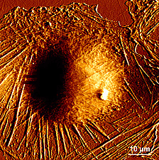
Living fibroblast cell image obtained by AFM

Force mode

In the force operating mode, the tip oscillates vertically while the cantilever deflection is registered.
This method has as its main applications the examination of specific interactions between molecules (antigen-antibody, the complementary strands of DNA), structural interactions (protein folding) or the study of polymer elasticity.


[1] This law states that the lenghtening of materials is directly proportional to the applied force on the same.
[2] Mechanical devices whose function is provide force to move another mechanical device.

Sources: http://www.dme-spm.com/funktion.html
              https://es.wikipedia.org/wiki/Microscopio_de_fuerza_at%C3%B3mica
              http://www.phy.mtu.edu/nue/images/atomicforce/Nanoscope2.jpg
              http://slideplayer.com/slide/6856881/  
              http://www.biozentrum.unibas.ch/research/groups-platforms/overview/unit/lim/


Read more

1.3.6. Repurposing drugs

0 comments
So far, we have studied the basics concerning the drug discovery process.Diversos tipos de fármacos
We have seen how these discoveries were made, at the beginning, by serendipity to give way later to the methodogical method: the target-based approach.
We still do not know how the new drugs will be discovered in the future, but biologics, genetics and phenotypic[1] screening in drug discovery will increasingly play a critical role.

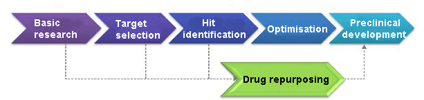
There is another area of increasing interest within the drug development pipeline that, although it does not belong to the discovery process, should be remarked upon. It is usually named drug repurposing, and it is based on giving a known compound a new use.
This practice consists of choosing a compound from the catalogue of shelved drugs that have failed in their clinical trials, or even have been commercialised, for the purpose of treating a different disease.

The main advantage that pharmaceutical developers find in reexamining these known compounds is that their safety profile and chemical properties are already well studied and understood, which considerably reduces the chances of failure in later stages during the development process.
Stage of repurposing drugs during the development process

Existing drugs can be repurposed in two fundamental ways:

The first one is to use them for a new or different therapeutic target. Sometimes drugs are shelved because they are considered too “dirty”, i.e. they bind too many targets and give various undesirable side effects. It may be that a different dosage or route of administration can reach a new target, and hence, be used to treat other pathologies.

The second method is based on taking a known target and a known drug for treating a different disease. We can find an illustrative example of this in the medication called finasteride, which originally was developed against prostate enlargement, and later it was discovered that it could stop or even reverse male pattern alopecia.
In spite of the fact that this medication performs on the same target for both pathologies, its approval to treat male baldness was eight years later than the authorisation for prostate enlargement. This was due to the fact that finasteride had to pass successfully a series of clinical trials before receiving its permission to treat the second indication.
Approaches for drug repurposing

A medication that fulfills both types of repurposing is chlorpromazine, which was originally produced as a antihistamine[2] and later was used as an antipsychotic on account of its sedative effect. While, these are very different illnesses, the drug acts on the same target for both.
Currently, chlorpromazine is used in combination with chemotherapy since its anticancer potential was discovered some years later. This property was learnt thanks to its ability to bind a previously unknown target.
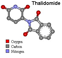
Thalidomide molecular structure
But the medicine that is many times seen as model of repurposing drugs is thalidomide. In principle, this medication was developed to treat morning sickness[3] in pregnant women.
When this drug was approved, the safety regulations were not as rigorous as they are today, and some years after it was found out that thalidomide was responsible for serious birth defects; 20,000 people were affected worldwide.
As a consequence, this drug was removed from the market for 50 years until the company Celgene studied it again, in order to use it against leprosy and multiple melanoma. After this new research, this medication was authorised again, warning strictly against its use by potentially pregnant women.


[1] Phenotype: set of an organism's visible traits such as eye and hair color, height...It is the interaction between a genotype and the environment. 
[2] Drug used to reduce or eliminate allergy effects. 
[3] Nausea in pregnancy, usually in the first few months.

Sources: UTAustinX: UT.4.01x Take Your Medicine - The Impact of Drug Development.
              http://www.imujer.com/salud/4707/tratamientos-naturales-para-la-enfermedad-de-la-manana
              http://www.academica.mx/observatorio/noticias/desarrolla-la-unam-f%C3%A1rmaco-controlar-algunos-tipos-c%C3%A1ncer
              http://www.elsevier.es/es-revista-educacion-quimica-78-articulo-avances-el-diseno-farmacos-asistido-90434983  
              http://www.dddmag.com/articles/2007/09/innovative-strategies-drug-repurposing
              https://embryology.med.unsw.edu.au/embryology/index.php/Abnormal_Development_-_Thalidomide


Read more

2.2.3. Nanobiosensors

0 comments
The great potential that nanotechnology offers enables the progress of new devices in the nanodiagnostic field, mainly in nanobiosensors.
Their properties are characterised by the nanoscale at which they are produced. Unlike biosensors , which we studied in the post: "Introduction to biosensors", where their sensory receptors are distanced by hundreds of micrometers, here they are distanced by a few nanometers.
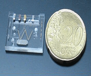
Actual size of a lab-on-a-chip
Thus, even sensors constituted by single molecules bound to their surface have been designed. Consequently the process of diagnosis is performed with a much smaller size device, improving portability and the potential for use anywhere.
Also, only extremely small sample quantities (micro or nanolitres) are required to carry out the analysis, so the sample extraction methods are less traumatic and invasive for patients.

These instruments also facilitate the use of samples without fluorescent or radioactive markers, which are used in biological and clinical analysis. This property is essential, since it has been proven that when a sample is not marked or altered before examining, the detection sensitivity significantly increases.

Within nanobiosensors we can distinguish the following categories:


Nanophotonic biosensors

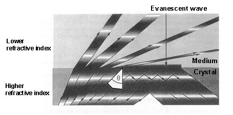
This class of nanobiosensors, also known as evanescent wave biosensors, is based on how the light is transmitted by multiple internal reflections through the optical waveguide, which is a component of the biosensor.

Evanescent wave productionWhen light is propagated in a medium and passes into a lower refractive index[1] medium, it is not totally reflected. As a consequence, a light component (called evanescent wave), in each of these reflections, is propagated in the lower refractive index that covers the waveguide. This propagation is really short, a few hundreds of nanometers, but it permits interaction between the light and the detection surface of the sensor, demonstrating which specific bioreceptors anchored to that surface interact with the analysed sample.

With these devices, only a few microliters of sample are necessary to determine protein concentrations or the variations of just one DNA base.

Also, it is possible to evaluate the metabolic state of just a single cell because some of these nanobiosensors have sharp fibre optic (30-50 nm) filaments, which can be introduced into cells through the plasma membrane without harming the cell.
This method facilitates the study of in vivo cell functions (apoptosis[2], cell division, biological nanomachines…) and it can detect pathological changes in a single cell.


Nanoplasmonic biosensors

In the Surface Plasmon Resonance (SPR) biosensor, a thin metal layer (normally a 50- nm-thick layer of gold) is placed on a dielectric material (a crystal), and its functioning is based on the detection of changes in the refractive index around the separation area between both elements.
By exciting the interface between these two layers (under conditions of total internal reflection) a plasmonic resonance[3] is activated with a certain angle of incidence.. This angle, which produces a plasmonic wave (with an evanescent character) is very sensitive to the changes from molecular interactions that happen on the metal surface. Therefore, these interactions between the analyte[4] of the sample and the sensor surface of the biosensor are shown as variations of the resonance angle.
Generation of Surface Plasmon Resonance (SPR)

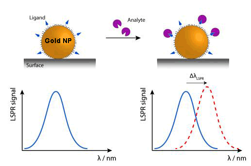
Currently, new biosensors are being developed, which rely on the plasmon resonance phenomenon in nanoparticles. While their detection range would be very similar to the SPRs, their functioning system can be made simpler by measuring light transmission instead of light reflection, in addition to the advantage of the device miniaturisation.

Due to the tiny size of nanoparticles, the electron oscillations are more localised than in the previous case (SPR), in specific areas of nanoparticles. This phenomenon is called ‘Localized Surface Plasmon Resonance’ (LSPR). The colour (wavelength: λ) changes that nanoparticles experience by adsorbing[5] the (bio)molecules of the examined sample are used to analyse this sample.
Variation of wavelength (Δλ) in nanoparticles when LSPR is produced

An alternative, within this kind of device,, is DNA sensors detecting colour changes produced by the aggregation of gold nanoparticles marked with stranded DNA complementary to the target DNA.

Nanoplasmonic biosensors are characterised by their real-time detection, specificity, high sensitivity and recognisition rate.
Their main applications are found in veterinarian, biomedical and environmental fields as well as in the food industry.


Nanomechanical biosensors

These sorts of nanobiosensors use the deflection or the resonance frequency variation of a microcantilever (as a transducing[6] method) when they interact with the studied sample. This position variation (Δx) is just a few nanometers derived from the biomolecular identification of the analysed sample.
Working principle of nanomechanical biosensors

Because these microcantilevers can be produced in mass at a low cost by standar microelectronic technology, thousands of them can be manufactured to identify thousands of analytes in a single sample.

The sensor area of these microcantilevers is around 1,000 μm2, which provides them to test sample volume lower than femtomole (10-15 moles).

Both photonic and nanomechanical biosensors could help us to obtain unlimited genetic and proteomic information that would offer the discovery of pathogens, new drugs, vaccines and undiscovered mutations of certain diseases in a faster way than the current technology.


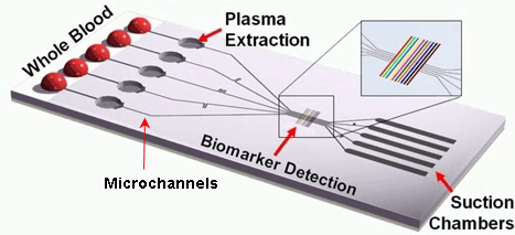
Microfluidic devices or lab-on-a-chip (LOC)

This class of instruments uses an electric field to move liquids, particles, molecules or cells through microcapillaries designed on chips of different materials like silicon, crystal, quartz or plastic.

A schematic diagram of a  lab-on-a-chipThis kind of chip is configured by a large number of integrated microchannels and microchambers, where the examined sample is subjected to complex chemical and biochemical reactions.
The required volume of the sample fluids is very small, therefore the analysis can be carried out very quickly.
It is a portable and disposable detection method with a high grade of automation.

The ultimate objective for all these biocompatible nanodevices is to finally be implanted in vivo inside our organism, where they would perform a similar function to ‘sentinels’ in the presence of the first diseased cells. Some advances have already been made to that effect at the microscale (pills with built-in image cameras), but, without a doubt, this will become one the major research fields in nanomedicine in the near future.


[1] It describes how light propagates through a certain medium. It is defined as n = c/v, where c is the speed of light in vaccum and v the speed of light in that medium.
[2] Process of programmed cell death.
[3] Excitation or oscillation of conduction electrons at the interface between two materials, in our case a metal layer on a dielectric substrate.
[4] Substance (ion, compound or element) of interest in an analysed sample.
[5] Adhesion of atoms, ions or molecules to a surface.
[6] Process by one signal or energy is converted to another.

Sources: Nanomedicina: aplicación de la nanotecnología en la salud. Laura M. Lechuga. Grupo de Nanobiosensores y Aplicaciones Bioanalíticas
              Centro de Investigación en Nanociencia y Nanotecnología (CIN2). CSIC
              http://www.kennislink.nl/publicaties/de-opmars-van-de-twentse-lab-on-a-chip
              http://images.slideplayer.es/16/5040279/slides/slide_47.jpg
              http://www.mdpi.com/sensors/sensors-10-09630/article_deploy/html/images/sensors-10-09630f1-1024.png  
              https://www.ifm.liu.se/applphys/molphys/research/biosensing_using_nanopart/
              http://www.tcd.ie/Physics/people/Martin.Hegner/ReviewNSST-The_impact_of_STM_and_AFM.html?ntherodt_01.pdf
              Fritz, J., Baller, M.K., Lang, H.P., Rothuizen, H., Vettiger, P., Meyer, E., Guntherodt, H.-J., Gerber, CH. and Gimzewski, J.K., 
              Science 288 (2000) 316.
              https://www.theengineer.co.uk/lab-on-chip-device-promises-hiv-diagnosis-in-10-minutes/


Read more

3.4. Proteins

0 comments
Proteins are the biological macromolecules with the largest variety in structure and functions. We can find thousands of them in a single cell, each performing a unique and specific role. In this way, we find proteins carrying out transport or storage functions, protective, contractile and structural proteins, enzymes or toxins.
All of them have in common that they are made up of structural polymers of amino acids arranged in lineal chains.


Types and functions of proteins

      Enzymes: they are known as complex or conjugated proteins, whose essential function is to act as an organic catalyst in biochemical reactions, where they assist to synthesise, reorder or break down different elements on which they perform.
An example are digestive enzymes, like salivary amylase, hydrolyses amylose (starch component).
      Hormones: they control and regulate physiological processes such as metabolism, growth or reproduction. They are chemical-signalling molecules, usually stereoids or small proteins, released by endocrine cells.
Examples include insulin, which is a protein hormone in charge of regulating glucose levels in blood.

Proteins present a wide range of molecular weights and shapes, where shapes are vital to their activities. But these functions can be modified by the exposure to chemicals and changes in pH or temperature, which lead to shape variations, and as a consequence the loss of their functions in a process named denaturation.


Amino acids

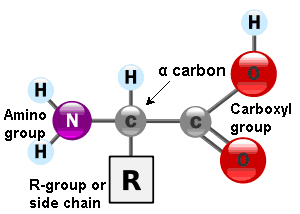
Amino acids are the monomers that constitute proteins. Each one has a structure formed by a central carbon atom, known as α carbon, to which an amino group (NH2), a hydrogen atom and a carboxyl group (COOH) are attached. The name ‘amino acid’ comes from the presence of these two groups in the amino acid basic structure.
General structure of amino acids
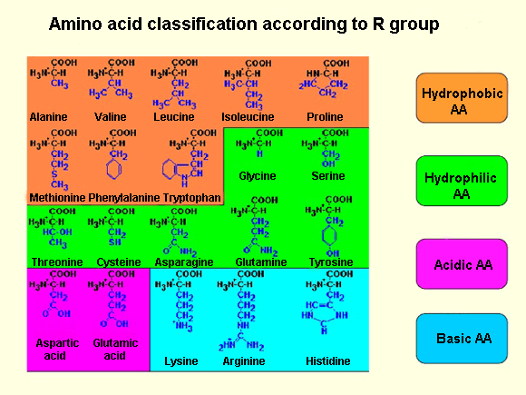
Each amino acid also have another atom or group of atoms joined to the central atom, called R group or side chain which is different for each kind of amino acid. The chemical nature of this chain establishes the type of aminoacid (whether it is polar, nonpolar, basic, acidic…). Thus, we can find basic aminoacids, like arginine and lysine, with a positive charge. Others such as cysteine or serine are polar due to their hydrophilic side chains, or if this chain is hydrophobic we have nonpolar amino acids like alanine or valine.
Amino acid classification according to R group
Out of the twenty different amino acids that form proteins, ten of them are considered essentials, because they are the necessary blocks for the protein construction. However, the human body cannot sinthesise them and must be obtained from the diet.

Amino acids are represented by a single upper case letter or three-letter abbreviation, e.g.: cysteine is symbolised by the letter C or the three letters Cys.

The number and sequence of amino acids ultimately set up the shape, function and size of proteins.

Amino acids are linked by covalent bonds, called a peptide bond, in which the carboxyl group of one amino acid is bonded to the amino group of the adjacent amino acid, releasing a water molecule during the process (dehydration reaction).
These bond products are called peptides and their joining give rise to a polypeptide chain. Each one has a free amino group at the end of the chain, named N terminal or amino terminal, having a C terminal or carboxyl terminal at the other end.

Often, the terms polypeptide and protein are used interchangeably, although technically a polypeptide is an amino acid polymer, while a protein is a polypeptide or combined polypeptides, each one with distinct shape and unique functions.


Protein structure

As I previously mentioned, protein shape is vital for the role they perform. In order to understand how a protein adopts its shape or final conformation, it is necessary to study the four structural levels in which they are organised: primary, secondary, tertiary and quaternary structures.

Primary structure
The primary structure is defined as the unique sequence of amino acids in a polypeptide chain. For example, the insulin hormone has two polypeptide chains, A and B, formed by unique sequences of 21 and 30 amino acids respectively and joined by two disulfide bonds between the cystein amino acids. There is also a third disulfide bond in the A chain, between two cysteine amino acids, which enables the molecule to fold into the suitable shape.

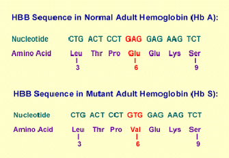
The unique sequence of every protein is ultimately determined by the genetic coding of that protein. Any alteration in the nucleotide[1] sequence, which constitutes the gene, can produce different amino acids that will be, later, added to the polypeptide chain and consequently affect the activity and structure of the resulting protein.

Thus, a single change in one of the 600 amino acids that make up the haemoglobin molecule origins, such a variation in its structure and activity, produces the appareance of sickle cell anaemia[2].Specifically, in the β chain of this protein, glutamic acid is substituted by valine.
More precisely, since every amino acid is made up of three nucleotide bases, those 600 amino acids generate 1,800 bases and sickle haemoglobin, the cause of sickle cell anaemia, arising from a single mutation in those 1,800 bases.
Mutant adult haemoglobin
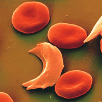
Sickle cells
As a result of this, haemoglobin molecules form long fibres that deform the disc or biconcave shape of red blood cells, acquiring a sickle or crescent shape that blocks arteries. This obstruction causes a series of disorders such as headaches, abdominal pain, diziness or breathlessnes so typical of this sickness.

Secondary structure
Secondary structures are generated from the local folding of polypeptides in certain regions, the most common are the α-helix and β-pleated sheet structures.
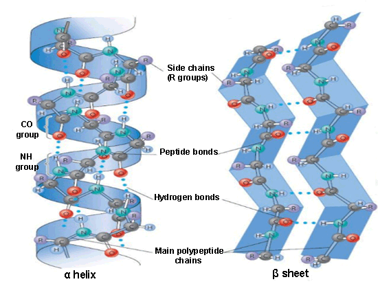
Secondary structures: α helix and β sheetThe α-helix is formed from the primary structure by rolling and twisting helically, with 3.6 amino acid residues per turn. They are constituted by hydrogen bonds between the carbonyl group (C=O) of one amino acid and the amino group (N-H) of another amino acid located four positions further in the polypeptide chain.

In the beta formation, amino acids are arranged in a zigzag way, named the ‘pleated sheet’ arrangement. Also it is originated by hydrogen bonds between N-H and C=O groups, but in this case they belong to adjacent chains.

Tertiary structure
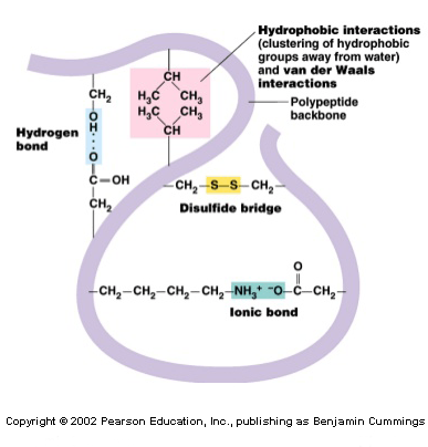
The tertiary structure informs how the secondary structure of a polypeptide folds up over itself giving rise to a globular conformation.Main bonds of tertiary structure
This configuration determines the physicochemical properties of proteins and assists their solubility in water, enabling, this way, it to carry out hormonal enzymatic and transport functions. This structure retains its stability due to the bonds among the R groups of amino acids. These bonds are created from the following types of interactions: disulfide bridges[3], hydrogen bonds, ionic and hydrophobic interactions and Van der Waals forces (to refresh all these concepts, visit the post titled: “Chemical bonds”).
In this arrangement, the nonpolar amino acids are, generally, facing the inside of the protein and the polar amino acids facing the outside, interacting with the surrounding aqueous medium.

Quaternary structure
In nature, some proteins are formed from several polypeptides, known as ‘subunits’, and their interaction creates the quaternary structure. These are weak interactions that assist the stabilisation of the overall structure of the protein.
The quaternary structure, usually, gives the protein its function and also creates many crucial biological structures, e.g.: viral capsids[4], microfilaments[5], microtubules[6] and collagen fibres of the connective tissue.
Structural levels of proteins


Denaturation and protein folding

Every protein has its own sequence and shape that are maintained through chemical interaction.

If the protein is exposed to chemicals or subjected to variations in pH or temperature, then its shape and structure can be modified without affecting its primary structure. This process is called denaturation, which can be reversible as long as the polypeptide primary structure is invariable during the process, allowing the protein to maintain its activity.
On the other hand, when the process is irreversible, the protein loses its function. This is what albumin (protein of egg white) experiences by being cooked, since it is denatured when it is exposed to high temperatures.

Protein folding plays a vital role for protein functions. This folding is made with the help of a protein family, named chaperones (or chaperonins), present in all cells. They are not part of functional proteins[6], but they bind to them to assist in their folding, their assembly and their cell transport to other parts of the cell where the protein performs its activity.


[1] Monomers of nucleic acids (DNA and RNA).
[2] Crescent or disc-shaped structures.
[3] Strong covalent bond between thiol groups (-SH) in the cysteine amino acid.
[4] Protein coat of virus that protects the viral genome.
[5] Thin fibres of proteins which with microtubules form the cell structure.
[6] Tubular structures of cells. Main component of cytoskeleton in eukaryotic cells.
[7] Those proteins that have a biological activity.

Sources: OpenStax College, Biology. OpenStax College. 30 May 2013.
              http://www.profesorenlinea.cl/Ciencias/ProteinasEstruct.htm
              http://study.com/academy/lesson/proteins-iv-higher-order-structure.html
              http://quimica.laguia2000.com/conceptos-basicos/cadena-lateral-en-aminoacidos
              http://es.slideshare.net/carolinacisdel/biomoleculas-enfermera
              http://biologiavfe.blogspot.com.es/2010/04/anemia-falciforme.html
              http://sicklecellcurefoundation.org/living-with-sickle-cell-disease/national-sickle-cell-month
              http://www.slideshare.net/VijayP7/secondary-structure-prediction-of-proteins
              http://www.slideshare.net/thelawofscience/protein-structure-11543259
              http://www.mobi4health.ug.edu.pl/wp-content/uploads/2014/10/2ndMSW-Milla-Neffling-Primer.pdf


Read more