• Biology

    Biology

    The atom, biological macromolecules (carbohydrates, lípids, proteins, nucleic acids), cell biology, cancer

  • Drugs

    Drugs

    Drug development, pharmacodynamics, pharmacokinetics, toxicology

  • Nanomedicine

    Nanomedicine

    Introduction to nanotechnology, diagnostic devices, drug delivery, regenerative medicine

  • Miscellaneous

    Miscellaneous

    A more in-depth explanation of topics previously mentioned in the other sections

1.3.2. Routes of drug administration

0 comments
After gathering all possible information about the lead compound, researchers must now understand how to manufacture a drug product. This applies not only to the lead compound but also to other chemicals that favour its disolution in the gastrointestinal (GI) tract, help in the manufacturing process or protect it from degradation.

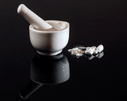
In addition to all these subtances, a wide array of products are added. In tablets, for example, cellulose is often included to improve the drug disintegration in the digestive fluids. Talc is also included in low concentrations during the manufacturing to absorb liquid and semi-liquid subtances. Lactose, or milk sugar, is often aggregated to bulk the tablet, acquiring a convenient size so that the patient can hold and swallow it.
Some tablets contain functional polymers to develop certain drug attributes. One of the main uses of functional polymers is to protect the tablet from the high acidity in the stomach, thus ensuring that the drug is dissolved in the small intestine later, and in this way improving its absortion in the intestinal fluids.

Even though a drug can be administered in many different ways, I will focus on those drugs that are administered orally to focus on the following questions:
Will the drug be dissolved and absorbed?, depending on how much it is absorbed, what should the dose be?, is it metabolised by the liver quickly after its absortion?, will it be degraded by the acidic media in the stomach?
With regard to the last question many drugs can be degraded at low pH.
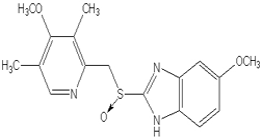
Chemical structure of Omeprazole
An example of these kinds of drugs is omeprazole, which without a functional polymer would not be effective. The purpose of this polymer is to preserve the omeprazole granules from direct contact with the gastrointestinal fluid until the pH becomes less acid and the drug is no longer in danger of being degraded. For this reason, it is important to remember that tablets and pills must not be broken, unless the pharmacist indicates it, or the polymer coating would be damaged and the drug would not be effective.
Routes of drug administration
Undoubtedly, the most usual way of delivering drugs to the body is orally because pills, capsules and tablets are easy to administer, mass-produced, have good stability and are easily absorbed by the digestive system.
But there are other systems that, at times, are better alternatives to the oral medication. Thus, intravenous or IV administration is performed when a high or well-controlled dose is required. This is the case with highly toxic agents (chemotherapy). However, this system has as a main disadvantage that is an invasive technique and it requires a clinician as well as sterile material to be dispensed.
Topical administration is another delivery system, for example ointments and creams to relieve irritations, rashes or insect bites as well as transdermal patches and gels which can permeate the skin so that the drug reaches the bloodstream.
Major routes of drug administration
A fourth type are inhaled medications, which like the former, perform locally, helping to treat illnesses like asthma, allergies or chronic lung diseases.Taking advantage of the fact that lungs are covered with a vast amount of vessels and capillaries, lots of drugs are inhaled (whether they are specific for the treatment of lung diseases or not) with the goal of being rapidly taken up by the circulatory system. As an example we can mention loxapine; an antipsychotic used in the treatment of schizophrenia, which when it is inhaled its effects can be detected in just ten minutes.

Therefore, the election of the suitable route of drug administration can make easier the drug uptake or even avoid possible side effects.


Sources: UTAustinX: UT.4.01x Take Your Medicine - The Impact of Drug Development.
              http://generalidadesdelafarmacia.blogspot.com.es/2010/11/vias-de-administracion-de-medicamentos.html
              http://www.xatakaciencia.com/medicina/farmacologia-vias-de-administracion-de-los-farmacos-y-sus-pros-contras              
              http://www.riesgoquimico.es/2009/03/30/omeprazol/


Read more
1.3.1. Pre-clinical phase

0 comments
Drugs
We have already studied in previous entries the long and specialised process that leads to the discovery and selection among thousands of candidates of the lead compound.

Next, I will explain the stage named pre-clinic phase, which is developed between drug discovery and clinical trials. This stage is divided into two parts: one focused on determining the lead compound’s safety and the second based on incorporating the pharmacological complex in a drug delivery system. There are a wide variety of drug delivery systems, all of them with development, manufacturing, storage and use specific rules. Among the most common and popular ones we find oral medications (pills, capsules, tablets...).
Fases en el desarrollo de fármacos
Resuming where we left off in the last entry, we start from an optimised lead compound, which: is capable of binding to one of the therapeutic targets which play a major role in disease progress, has a structure that looks druggable and is apparently non-toxic.
From now on, the pharmaceutical formulator is in charge of defining the best way to administer this compound to its target or ‘action’ site within the organism.
The most effective methodology in support of this process is to keep concentrating on obtaining a broader knowledge of the compound itself, always keeping in mind the type of drug delivery system that will be used. To do this, we must ask a series of questions about its physical and chemical properties: is it soluble in water, in oil or a combination of both?, can it crystallise?, can these crystals reduced in smaller particles?, is it a stable drug from a chemical point of view?, if it is so, how long?, how is it degraded?, does it do in an acidic environment? All these questions must be answered before it is incorporated in an actual dosage form.

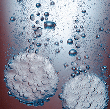
Dissolving aspirine
One of the most basic features that must be known about a drug is its solubility in water. When drugs are given most of them are in contact with some water-based biological fluid since the human body is mostly made up of water.
Solubility is a vital characteristic for the vast majority of drugs, but especially for those that are absorbed in the gastrointestinal (GI) tract. Thus, for example, an aspirin could not be absorbed in the small intestine even if it were split up into smaller fragments if it were not dissolved in the intestinal fluids.

In the case that a compound has no, or low solubility in water, the pharmaceutical formulator will try to increase its solubility. As a second alternative, a step back can be taken in this process and the compound can be sent back to a chemical laboratory where a salt form of the drug is synthesised. The compound solubility not only must be tested in aqueous media, but also in diverse acidic media, due to the fact that gastrointestinal fluids have diverse acidity levels.
In addition to water other non-aqueous solvents used in the final formulation of the drug must be evaluated such as glycerol, ethanol or propylene glycol, as well as solvents used in the manufacturing process like isopropyl alcohol, methanol and acetonitrile.

Sources: UTAustinX: UT.4.01x Take Your Medicine - The Impact of Drug Development.
              http://indacea.org/desarrollo-de-medicamentos-1/               
              Valentia Biopharma S.L.               
              http://www.pharmatechespanol.com.mx/articulo/820.polimeros_avanzados_para_mejora_de_la_solubilidad


Read more

2.1. Scanning tunneling microscope (STM)

0 comments
In the post titled “Nanotechonology. Introduction” I mentioned that the development of the scanning tunneling microscope played a fundamental role in the progress of nanotechnology, so let’s learn a little more about it and how it works.
The scanning tunneling microscope (STM) was developed by Gerd Binning and Heinrich Rohrer in 1981 at IBM laboratories in Zurich (Switzerland), for which they were awarded the 1986 Nobel Prize in Physics.
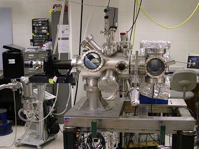
Scanning tunneling microscope
Both the STM and the AFM (atomic force microscope) are classified as scanning probe microscopy instruments, but the first is far more powerful and is able to handle and observe atoms and molecules with higher resolution.

The STM is used to take images of conductive surfaces at an atomic scale of around 2 Å (2*10-10 m). It can also modify the examined sample by manipulating individual atoms, setting off chemical reactions and originating ions by replacing electrons from certain atoms with others.

The scanning tunneling microscope is a non-optical microscope (it does not use rays of light to take us down to nano-dimensions) whose operation is based on quantum mechanic principles. An extremely fine probe is positioned over the specimen under study at a distance of the diameter of an atom, applying a voltage between them. Depending on the characteristics of that voltage, the electrons can jump from one side to the other by tunneling effect[1], producing a weak current flow, known as “tunneling current”, whose value is approximately a few picoamperes (1 pA = 10-12 A).
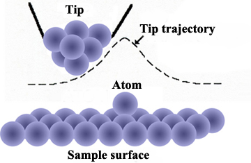
Trajectory of the probe tip over the sample
The stylus probe is very sharp, with a tip formed by a single atom. It scans the surface at a very slow speed, raising and lowering (by a piezoelectric mechanism[2] to control its level) in order to maintain a constant distance and signal, which permits the inspection of the tiniest detail of the sample that is being scanned.
The vertical movement of the stylus is registered allowing the inspection of the surface structure atom by atom, producing a contour map of the surface generated by a computer.

Electrical insulating materials cannot be analysed by this technique because, as their electrical charges do not flow freely it consequently makes it impossible to conduct any kind of current between them and the probe tip.

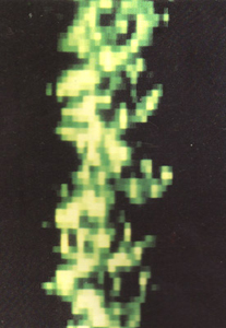
Parallel double-stranded DNA. Image obtained by STM
Although these instruments show an optimal operation examining conductive materials, they can also provide topographical characterisations of organic molecules such as DNA or proteins.

This type of microscope plays an important role in physics, specifically in the study of  semiconductor surfaces and in the field of microelectronics. It also proves to be important in chemistry, studying superficial reactions like catalysis or in nanoscale chemistry laboratories, where the analysis of the physical structure of synthetic chemical compounds and material defects is essential.


[1] The tunnel effect consists in those quantum particles (electrons in this case) that in spite of the fact of not having enough kinetic energy (energy associated with body motion), they penetrate and cross a space that, in principle, would be impossible, due to the presence of a potential barrier that should block their flow.
[2] Materials that are deformed through expansion or contraction, under the action of an electric field.

Sources: http://education.mrsec.wisc.edu/130.htm              
              http://www.nobelprize.org/educational/physics/microscopes/scanning/
              http://hoffman.physics.harvard.edu/research/STMintro.php
              http://www.uwec.edu/Matsci/center/instrumentation/


Read more

2.1. Introduction to nanomedicine

0 comments
The application of nanotechnology in the healthcare field has enabled the development of a new scientific discipline known as nanomedicine.

Due to the fact that some of the fundamental biological macromolecules of living organisms (like the DNA and proteins) are located at nanoscale, this has allowed the interaction between nanometric devices and nanomaterials within the human body. This has facilitated the discovery of important new advances in medicine.

Certain predictions made in the early stages of the development of nanotechnology are still science fiction.
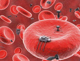
Imaginary nanobots in the bloodstream
For instance, the idea that we could develop “nanobots” that would protect us against external microorganisms, would cure injuries and damaged tissues selectively. Nevertheless, some major advances have been made.

The steady increase of neurodegenerative and cardiovascular diseases, diabetes or cancer  requires us to investigate new diagnostic and therapeutic techniques that must be simpler, quicker and more precise than the ones we have today, decreasing, at the same time, the costs involved. It is expected that nanomedicine should address some of these problems, like diagnosing disease at its earliest stage, tailored treatments for patients or the ability to regenerate damaged organs and tissues.

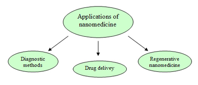
Nanomedicine is focused on three large areas: diagnostic methods, drug delivery systems and regenerative medicine. Nanodiagnostic techniques are divided into analysis systems and methods of medical imaging. Both methods enable the detection of the appearance of the disease, in vitro and in vivo at early stages.  This makes it possible to take action with the most appropiate treatment and causing as little damage as possible to the rest of the body.
Fields of applications of nanomedicine
In sample analysis of patients, nanomedicine also helps to achieve a more specific, quicker and more effective diagnosis. In this way diseases are diagnosed at cellular and molecular level providing more chance of a cure.
Nanomedicine has also advanced drug delivery systems: drugs are guided specifically to those affected areas and cells to accomplish a more effective treatment with a lower dose and minimising side effects. In addition, these systems protect the carried drug by avoiding degradation of the drug before it reaches its target. Thus, the challenges of  delivery associated with drugs with poor solubility or with those that cannot be delivered using conventional methods is solved.
Lastly, through gene and cell therapies, biomaterials, tissue engineering and nanotech tools, regenerative nanomedicine boosts the natural repairing mechanisms of the human body which often are not enough to achieve the total recovery of the organism alone.


Sources: Informe de vigilancia tecnológica: nanomedicina. Fundación para el conocimiento madri+d CEIM
              Nanomedicina: aplicación de la nanotecnología en la salud. Laura M. Lechuga.Grupo de Nanobiosensores y Aplicaciones Bioanalíticas
              Centro de Investigación en Nanociencia y Nanotecnología (CIN2). CSIC
              Fundación española para la ciencia y la tecnología (FECYT). Nanociencia y nanotecnología. Entre la ciencia ficción del presente y la                     tecnología del futuro, 2009.


Read more

3.2. Carbohydrates

0 comments
Most people are more than familiar with the first type of biological macromolecules that I will explain: carbohydrates.
Carbohydrates are an essential part of our diet; they provide energy (4.3 Kcal/g) mainly through glucose. Among the rich natural sources of carbohydrates we find: vegetables, fruits and cereals. Their molecular structure is represented by the stoichiometric formula (CH2O)n, where “n” indicates the number of carbon atoms in the molecule. Therefore, in carbohydrate molecules the ratio C, H, O is always 1:2:1.
Carbohydrates are divided into three types: monosaccharides, disaccharides, and polysaccharides.


Monosaccharides

Monosaccharides (mono-: one; sacchar-: sweet) are simple sugars; glucose being the most common among them. Most of them are named by the suffix: “-ose”. They contain a number of carbon atoms that vary from three to seven. Thus, we find trioses (three carbons), pentoses (five carbons) or hexoses (six carbons).
Monosaccharides are molecules that can adopt both linear chain shapes and ring shapes; these ring forms are frequently found in aqueous solutions. If monosaccharides contain an aldehyde group[1], the monosaccharide is known as aldose, however if the functional group is a ketone group[1], the monosaccharide is called ketose.

The monosaccharide glucose (C6H12O6) is one of the basic energy sources for the human being. During cell respiration, glucose releases energy which is used to produce ATP (adenosine triphosphate)[2]. Plants synthesise glucose using CO2 and water, their surpluses are stored/accumulated as starch which is catabolised (broken down by cells) when it is consumed by the human being or herbivorous animals.

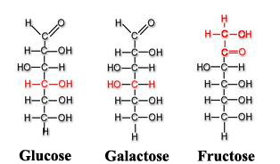
Among other monosaccharides, we can also point out galactose (which is part of lactose or milk sugar) or fructose (present in fruits and honey). Both glucose, galactose and fructose[3] are examples of isomeric monosaccharides because they have the same chemical structure but they differ structurally and chemically due to the different arrangements of their functional groups around their assymetric (or chiral) carbons[4]. Glucose and galactose belong to the group that we saw previously called aldoses and fructose belongs to the ketose group.
Glucose, galactose and fructose molecules

Disaccharides

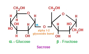
Disaccharides (di-: two) are formed when two monosaccharides undergo a dehydration synthesis. During this process the hydrogen atom of one monosaccharide is combined with the hydroxyl group of other monosaccharide, forming a covalent bond between them and releasing a water molecule. This covalent bond is named glycosidic bond, which is classified in two types: α and β, depending on if the hydroxyl group is located above or below the chiral carbon (also known as asymmetric carbon that we saw previously).
Sucrose molecule: combination of monosaccharides glucose and fructose
Among the most common disaccharides are found lactose, which is the result of the union of galactose and fructose. Milk is a natural source where lactose is found.  Another example of disaccharides is maltose, or malt sugar, which is formed during a dehydration reaction performed by two glucose molecules. But, without a doubt, the most common disaccharide is sacarose, or table sugar, made up of fructose and glucose monomers.


Polysaccharides

Polysaccharides (poly-: many) are long chains of monosaccharides joined by glycosidic bonds. These chains can be branched or unbranched, containing different types of monosaccharides.
Among the main polysaccharides are: cellulose, glucogen, starch and chitin.

Starch
As I mentioned earlier, plants are capable of synthesising glucose from water and CO2. When glucose production exceeds the energy needs of the plant, the surpluses/excesses are stored as starch in different parts of the plant like the root and seeds, providing feed during their germination.
When starch is consumed by animals or humans in their diets, it is first broken down by enzymes (such as salivary amylases) into smaller molecules like glucose, before being absorbed by cells to satisfy their energy needs.

Glucogen
Glucogen, composed of glucose monomers, is how glucose is stored in humans and other vertebrates. It is the equivalent to starch in plants and it is accumulated in muscular cells and the liver. When blood glucose levels decrease, these glucose deposits are demanded and glucogen is broken down into glucose during a process called glycogenolysis.
Glycogenesis and glycogenolysis processes

Cellulose
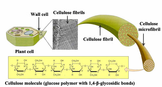
Cellulose, also made up of glucose monomers, is the most abundant polymer. It is the main element of plant cell walls, giving them structural support. In cellulose the basic monomers are tightly packed, giving cellulose its typical fibrous structure which confers stiffness and high tensile strength, i.e. structural support for plants.
Unlike human beings, herbivores have bacteria and protists[5] inside their digestive system which segregate the cellulase enzyme to help digest the cellulose they consume in their grass. By the action of this enzyme, cellulose is broken down into glucose monomers and in this way it can be used as energy source.
Cellulose structure

Chitin
The fourth polysaccharide to point out is chitin, which is the main component in fungi cell walls and the exoskeleton (external skeleton) of arthropods, which protects their internal organs. Chitin is a polysaccharide with a high nitrogen content, constituted by repetitive units of N-acetyl-β-glucosamine, a kind of modified sugar.


BENEFITS OF CARBOHYDRATES

Carbohydrates are composed of both soluble and insoluble elements. Among the insoluble carbohydrates we find fiber, which is mainly cellulose. Cellulose helps to regulate the intestinal transit and the glucose consumption rate. It also binds to the blood cholesterol in the small intestine and in this way the cholesterol is eliminated by being excreted from the organism in faeces. Also, diets high in fibre can help to protect against colorectal cancer.

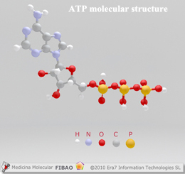
Glucose is provided by the carbohydrates that we consume. Once it is broken down during cell respiration, glucose produces ATP molecules, known as the energy-currency of cells, providing fast energy for any organism.
ATP molecular structure
Consequently, without the consumption of carbohydrates, the availability of instant energy for the body (or any other organism) would be deeply reduced.

For all these reasons, eliminating the ingestion of carbohydrates, like some hypocaloric diets recommend in order to achieve a fast weight loss, is questionably healthy. To accomplish such an aim, it would be more healthy to balance appropriate low GI carbohydrates present in vegetables, fruits and cereals with a balanced level of proteins, vitamins and lipids, a suitable water intake and doing exercise appropriate to our age, gender and physical fitness.


[1] Both are organic compounds characterised by containing the carbonyl functional group (C=O). But aldehydes contain the carbonyl group bonded to at least one hydrogen atom, whereas ketones contain the carbonyl group bonded to two carbon atoms.
[2] Molecule used by all living organism to provide energy in chemical reactions. It is considered as the “energy-currency” of the metabolism.
[3] They all are hexoses: six carbon atoms in their structures.
[4] Carbon atom attached to four different types of atoms or group of atoms.
[5] Eukaryotic (their cell nucleus is delimited by a membrane) organisms, usually unicellular, which due to their own characteristics cannot be included in the rest of the kingdoms (animals, plants and fungi) within this category.

Sources: OpenStax College, Biology. OpenStax College. 30 May 2013.              
              http://medmol.es/glosario/121012glosariomedmol_atp/
              https://kel-tay-lii.wikispaces.com/A.+Intro+to+Phys
              http://chemistry.tutorvista.com/organic-chemistry/oligosaccharides.html
              http://www.entrenasalud.es/glucogeno-y-deporte-un-gran-deposito-de-energia/
              http://www.asturnatura.com/articulos/envoltura-celular/pared-celular.php


Read more

3.1. Biological macromolecules. Introduction

0 comments
Within this section I will explain the four main types of biological macromolecules: carbohydrates, lipids, proteins and nucleic acids. These macromolecules are the basic constituents of cell structure, that in combining form most of the dry weight of a typical cell. They carry out a wide range of functions in cells. These macromolecules are organic molecules, thus their main component is carbon. Hydrogen (H), oxygen (O) and nitrogen (N) and other minor elements are also part of their structure.
The two fundamental reactions that control the synthesis of biological macromolecules are the following:

    ▣      Dehydration synthesis
The basic blocks of most macromolecules are sub-units known as monomers. These monomers are combined through covalent bonds to form larger molecules (polymers). To do this they release water molecules as byproducts, this mechanism is called dehydration synthesis, which means “to join by losing water”. During these reactions, the hydrogen (H+) of one of the monomers is combined with the hydroxyl group (OH-) of other monomer, releasing a water molecule. At the same time, the monomers share electrons and are joined by covalent bonds.
Different types of monomers or even just a single monomer is combined to form a diverse range of polymers. Thus, for instance, glucose monomers are the components of several different substances such as cellulose, glycogen or starch.
Dehydration reaction

    ▣      Hydrolysis
Hydrolysis (“to separate water”) is a reaction in which water molecules are used to break down the polymers into their basic monomers. In these reactions the polymers are divided in two: one part acquires a hydrogen atom (H+) and the other a hydroxyl group (OH-); both elements (H+ and OH-) occur from the breakdown of a water molecule.
Hydrolysis reaction
Both in hydrolysis and dehydration reactions, enzymes (a specific type of proteins) can act to catalyse (accelerate in these two cases) these reactions.
Through hydrolysis and using particular digestive enzymes, our body breaks down the proteins, lipids and carbohydrates (macromolecules) that we eat. Once these ‘macro’ (i.e. larger) molecules are broken down into smaller molecules, the intestinal cells can absorb the nutrients that they contain. Thus, for instance, lipids are metabolised by lipases, proteins are broken down by the action of hydrochloric acid, peptidase or pepsin and finally carbohydrates are digested by enzymes like lactase, maltase or sucrase.
By breaking down all these molecules into a unit where cells can absorb them as their own ‘food’, these cells obtain the necessary energy to perform all their functions.

Sources: OpenStax College, Biology. OpenStax College. 30 May 2013.               
              http://www.differencebetween.net/science/health/difference-between-hydrolysis-and-dehydration-synthesis/


Read more