4.5.2. Cell reproduction II

0 comments
The cell cycle is a set of ordered events that involve the cell growth and division to produce two new daughters cells.

Prior to cell division, cells go through a series of stages of growth, DNA replication and division, carefully regulated, with the objective of creating two identical cells.

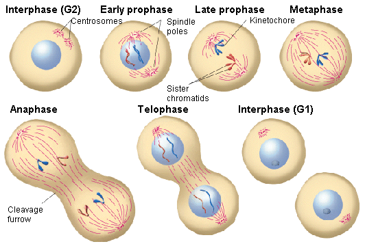
The two main phases of the cell cycle are the interphase and the mitotic phase. During the first stage, the cell grows and replicates its DNA. While, in the mitotic phase, the replicated DNA and the cytoplasmatic content come apart and the cell divides.


Interphase

The interphase is the longest period of the cell cycle, it occurs between two mitosis and we can distinguish the following phases within it:
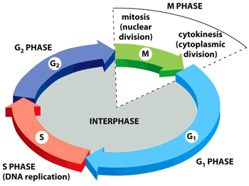
Interphase stages
      G1: the cell grows, reaching its critical size. RNA and proteins are synthesised and the centrosome is duplicated. If conditions are appropiate, the cell passes the restriction point (a G1 phase checkpoint) and the next step begins.
Differentiated cells that do not divide (e.g., neurons or cells from the cardiac muscle) leave the cell cycle at this point and start the G0 phase[1].
      S: DNA is replicated. In this way, two daughter cells inherit the same genome from the mother cell.
      G2: repair phase of DNA and preparation for mitosis. The cell doubles its size compared to G1 phase.


Mitotic phase

The mitotic phase is a multistep process in which the duplicated chromosomes align, come apart and move to the two new daughters created.
The first stage of the mitotic phase is called karyokinesis, mitosis or nuclear division and the second step is known as cytokinesis, which is the physical separation of the cytoplasmatic components into the two daughter cells.
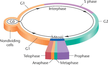
Mitosis, where the genetic material is divided equally, comprises the following stages:
Somatic cell cycle
      Prophase: chromatin is condensed, the mitotic spindle[2] is formed and the nucleolus disappears.
      Prometaphase: the nuclear envelope is removed and the chromosomes join the microtubules through the kinetochores[3].
      Metaphase: the chromosomes align randomly in a plane called metaphase plate, or equatorial plane, between the two poles of the cell.
      Anaphase: the microtubules of the spindle become shorter, which causes the separation of the two sister chromatids.
      Telophase: the chromosomes decondense and the nuclear envelope, along with the nucleoulus is formed. Lastly, the mitotic spindle disappears.

During the last step of the mitotic phase, cytokinesis, a contractil ring of actin filaments is produced which divides the cytoplasm into the two daughter cells.
Mitotic phase stages


Control of the cell cycle

The duration of the cell cycle is higly variable, even in cells of the same organism. In human beings, it ranges from just a few hours in the embryonic cells, to two - five days for epithelial cells, to a whole human life for specialised cells, like cardiac muscle cells or cortical neurons.

The time that a cell spends in each phase of the cycle varies too. Thus, for example, those human cells with a 24-hour cycle spend approximately nine hours in the G1 phase, ten hours in the S phase, around four and a half hours in the G2 phase and about 30 minutes in the M phase.

Each step of the cell cycle is controlled by mechanisms both internal and external to the cell.

Regulation of the cell cycle by external mechanisms
Both the start and the termination of the cell replication cycle are activated by events external to the cell.
For instance, events such as the death of a near cell, the release of hormones that favour growth or the cell reaching a specific size can start cell division.
Regardless of the type of externally received message, a series of internal effects then lead it to the interphase.
From this starting point, each required parameter in each phase must be satisfied so that the cell cycle can continue.

Regulation at internal checkpoints
The checkpoints of the cell cycle prevent mistakes in the duplication or in the distribution of chromosomes (which can cause mutations).
We can distinguish three checkpoints:
      The G1 checkpoint: also named the ‘rectriction point’ in mammals. It acts at the end of G1 phase and determines if all the necessary requirements are fulfilled to proceed the cell division.
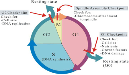
Internal checkpointsSi no se cumplen, la célula puede parar el ciclo hasta que las condiciones adversas se solucionen o puede pasar a la fase G0 y esperar señales que indiquen que las condiciones han mejorado.
      The G2 checkpoint: carries out its role during the transition between G2 to M phases. Its most important function is to make sure that the chromosomes have been replicated and that replication has been accomplished without DNA damage. If damage is detected, the cycle stops and the cell tries to complete the DNA replication or repair the damaged one.
      The M checkpoint: is also known as the spindle checkpoint. It acts near the end of the metaphase stage of karyokinesis. It determines whether the sister chromatids are firmly and correctly bound to the spindle microtubules by the kinetochores.

The control system of the cell cycle could be compared to the program of a washing machine, where the washing machine only carries out its functions when the signals from its sensors (lock door, levels of water and detergent, power supply) indicate that it is ok to move forward during the diverse stages of the washing cycle.


Regulator molecules of the cell cycle

In addition to the checkpoints, there are some intracellular molecules that regulate the cell cycle and can act individually or affect the activity or production of other regulator molecules.
Regulator molecules are classified into two groups:
Regulator molecules of the cell cycle (cyclins and CDKs)
      Those molecules that promote the progress of a cell to the next phase (positive regulation). They are, in turn, divided into two types: cyclins[4] and cyclin-dependent kinases (CDKs)[5].
      The second group are negative regulators, which are in charge of halting the cell cycle. The best-known negative regulators are retinoblastoma protein (Rb), p53 and p21.
Retinoblastoma protein is a suppressor protein for tumours that is altered in many kinds of cancer (prostate, breast), but it is retina cancer from where it takes its name.
P21 inhibits the cell cycle, with its levels controlled, in turn, by p53, which regulates cell growth and controls DNA damage. This protein can halt the cycle if needed, and is able to cause apoptosis too.


[1] Permanent or temporary quiescence (inactivity) phase in which the cell cycle stops.
[2] Set of microtubules whose function is to enable the migration and correct separation of the chromosomes during mitosis.
[3] Protein structures whose function it is to initiate, control and supervise the movement of the chromosomes during cell division.
[4] Proteins synthesised during the interphase and destroyed at the end of the mitosis. They regulate the enzymatic activity of CDKs.
[5] Enzymes that activate or inhibit other proteins by phosphorylating them (adding phosphate groups).

Sources: OpenStax College, Biology. OpenStax College. 30 May 2013.
              http://www.biocancer.com/journal/1219/41-regulacion-positiva-del-ciclo-celular
              http://www.pucmmsti.edu.do/websise/estudiante/materias/201120122/ST-BIO-112-T-003/04.%20Regulacion%20del%20ciclo%
              20celular%20Apoptosis%20Cancer%20%20.pdf
              http://bio3400.nicerweb.com/Locked/media/ch02/interphase.html
              http://www.slideshare.net/obessionforu/cell-cycle-and-molecular-basis-of-cancer
              http://biologicalexceptions.blogspot.com.es/2013_01_01_archive.html
              http://gleesonbiology.pbworks.com/w/page/7537859/C9
              http://www.bioscirep.org/content/30/4/243


Your opinion matters

4.5.1. Cell reproduction I

0 comments
Human beings, like other organisms that reproduce sexually, start life from a zygote or fertilised egg.
Later, trillions of cell divisions take place in a controlled way to lead to a complex multicellular human being, or in other words, a single cell is the common ancestor of the rest of the organism’s cells.
Even when a living being has developed completely, cell division is still necessary for the development, maintenance and repair of cells and tissues. This division is strictly regulated to avoid consequences that might be fatal for that organism.


Cell division

Cell reproduction via the cell cycle constitutes the foundation that enables the continuity of life from one cell to another. This cycle, strongly regulated, includes the diverse stages of a cell life from the division of a single parent cell to the reproduction of two new daughter cells.

Before going on with the cell reproduction process, it is necessary to understand the function and structure of the genetic information of a cell.Chromosome structure
The number of chromosomes[1] of eukaryotic cells varies depending on the species.
In human beings, body cells (somatic cells) have 46 chromosomes, while sex cells or gametes (eggs and sperm) have 23 each.
Somatic cells have two sets of chromosomes (2n[2]) in a configuration called diploid.
Gametes, that have a single set of chromosomes, are designated as 1n and they are known as haploid cells.

Genes (functional units of the chromosome) determine particular features by coding specific proteins, to represent variations in specific features. For instance, eye colour is a characteristic whose traits can be brown, blue or green.
These traits in an individual are established by the inherited genes from each parent.

If the DNA of those 46 chromosomes in a human cell unfolded from one end to the other, it would measure around 2 metres and its diameter just 2 nm. Taking into account that the typical size of a cell is about 10 μm, DNA has to condense itself to fit into the cell nucleus and must be quickly accesible so that genes can be expressed.

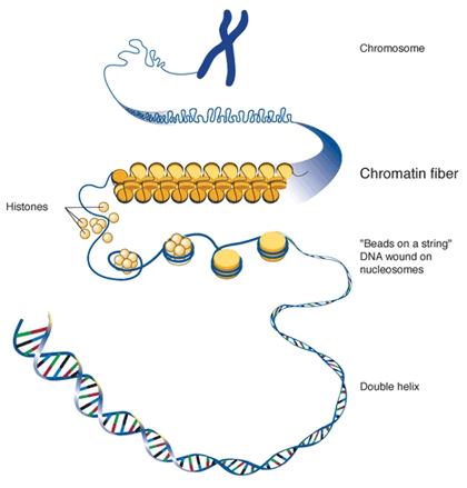
During several phases of the cell cycle, the long chains of DNA are highly condensed in the chromosome. There are a number mechanisms by which chromosomes are compacted, in all of them various classes of proteins help to organise and pack the chromosomal DNA.

In the first level of compaction, DNA is strongly packed around a core of eight structural proteins (histones) in regular intervals along the chromosome, forming the structure known as chromatin.
The fundamental unit of organisation for chromatin in eukaryotic cells is the nucleosome, composed of a DNA fragments around 200 base pairs (bp) long and a histone octamer (group of eight). This structure compacts seven times the DNA molecule which under a microscope looks like a ‘necklace of beads’ with a 10 nm diameter.
DNA condensation levels

In the second level of compaction, the nucleosome fibres and the linker DNA[3] roll into 30 nm fibres (DNA molecule is now 50 times packed).

The third level of compaction uses fibrous proteins to compact chromatin, creating radial loops of 250-400 nm long. These proteins make sure that each chromosome in the non-dividing phase occupies a certain area in the nucleus avoiding overlap.

In higher levels of organization we find chromatids (700 nm - 1μm long). When two of these chromatids join, through a region called the centromere, they form metaphasic chromosomes[4].


[1] Highly organized structures comprising DNA and proteins, which contain most of the genetic information of an individual.
[2] n indicates the number of chromosome sets.
[3] DNA regions that are not transcribed and are located between genes.
[4] Metaphase: second phase of mitosis (cell division) in which the nuclear membrane is removed and the chromosomes are placed in the equator of the cell.

Sources: OpenStax College, Biology. OpenStax College. 30 May 2013.
              https://www.quora.com/What-are-the-differences-between-chromosomes-chromatids-and-chromatin
              https://burningscience.wordpress.com/biology/dna-structure-and-replication/
              http://27.109.7.67:1111/econtent/cell-communication/principles-part-3.php
              https://www.quora.com/What-are-the-differences-between-chromosomes-chromatids-and-chromatin


Your opinion matters

1.6. Personalised medicine

0 comments
In recent years, the definition of a succesful drug has changed. The time when a drug would become a blockbuster is over, since there are no longer illnesses that can be treated with just a drug. At the same time, patients and markets demand more effective medicines.

But this demand turns out to be harder to fulfil because our knowledge about diseases is getting more and more complex. Besides, there are numerous external pressures (from governments, financial institutions, regulatory agencies and even scientists) to achieve the best clinical results.
Then, how is it possible to create new drugs when their development costs increase year after year and it is more difficult to generate them with a clinical benefit for patients?

To overcome this challenge, science has provided us with new tools, such as the ability to map the human genome (at a lower and lower cost), which allows us to understand the expression of different proteins in response to diverse illnesses.
Consequently, personalised medicine, which uses these new kinds of tools, is expected to take the lead in the discovery of new drug development processes and of specific medicines that are more effective and even safer.
Molecular testing in personalised medicine
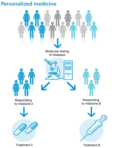
But, how is a personalised drug defined? Personalised medicine is that medical treatment tailored to the features of each and every patient.
It is expected that personalised drugs will be prescribed only to those for whom there will be a known and personal benefit.
In this way, not only the capacity to treat diseases improves, but also, the associated savings in prescribing in a targeted way.

Within personalised medicine, we find three categories:
      Diagnostics: these products can diagnose the risk of suffering from a specific disease, the real existence of a disease in an individual, or even guide the treatment for those people who have already been diagnosed.
      Companion diagnostics: : these products work in tandem with pharmaceutical treatments, indicating to the physician the type of drugs that a particular patient should receive.
      Screening tools: they are able to detect diseases in their early stages, which allows quick interventions with the hope of diminishing the effects caused by illnesses.

Ideally, it would be interesting to find a way of evaluating a person’s vulnerability to a disease or their response to a certain treatment even before administering it.
With the help of personalised medicine, we could know beforehand which people would respond positively to a specific drug and those that would not; which people would suffer grave side effects and those that would not; which people would need that medicine at an early stage and those who would need it at a later stage. All with the goal of accomplishing the best possible results in each patient.

Personalised medicine has experienced spectacular growth in the last few years, from 13 main products in the market in 2006 to 72 products in the year 2011 and sales of 30 billion dollars in the USA.
Presently, 94% of biopharmaceutical companies are researching personalised medicine. They are aware that it can be used both as a research tool as well as to improve a drug’s potential.
Tailored treatments of personalised medicine
Among the personalised medicine products that physicians use currently to treat their patients are the following:
      BRACAnalysis tests that identify potential mutations in the genes BRCA1 and BRCA2 responsible for most ovary and breast inherited cancers. Depending upon the presence or not of these mutations, the patient’s likelihood of developing these types of cancer in the future is determined. According to that, other relatives are guided and advised about what steps and decisions to make.
      Oncotype DX is a 21gene test that predicts a patient’s likelihood of benefiting from chemotherapy and the risk of a tumour recurrence in the following ten years.
      PreDX is a blood test that examines the levels of seven protein markers associated with diabetes. These markers are related to fat tissue quality, inflammatory conditions of the pancreas and glucose metabolism.

Some of the current benefits from personalised medicine include:
      A 34% reduction of chemotherapy in those women who suffer from breast cancer and prior to these treatments they have been undergone genetic testing.
      Up to 17,000 strokes can be prevented every year in those patients who are prone to cerebrovascular disorder by giving the proper dose of warfarin[1] provided that a genetic test is carried out.
      Over 600 million dollar savings for the health care system in the USA if patients with metastatic colorectal cancer undergo the KRAS gene[2] test before receiving their treatments.
      Development of new diagnostic tools that can help in the drug development process. Thus, if pharmaceutical companies know in advance what patients will respond better to a clinical trial, better results will be obtained in that study.

We are still in the early stages of personalised medicine, which will provide us with a better understanding about disease conditions, minimise side effects, and identify new drug targets.
By incorporating it into our experiments and research methods, we will also identify new innovations related to the drug discovery process.


[1] An anticoagulant (blood thinner) used in the prevention of thrombosis and thromboembolism.
[2] Gene that produces KRAS protein, which takes part in many cell signalling patways, cell growth and apoptosis.

Sources: UTAustinX: UT.4.01x Take Your Medicine - The Impact of Drug Development.
              http://www.patia.com.mx/braca.html
              https://breast-cancer.oncotypedx.com/es-MX/Professional-Invasive/WhatIsTheOncotypeDXBreastCancerTest.aspx
              http://www.tuorigen.cl/index.php/preguntas-frecuentes
              http://www.cancer.gov/espanol/publicaciones/diccionario?cdrid=652256
              http://www.lampadia.com/analisis/tecnologia/medicina-del-futuro-despliegue-de-un-nuevo-paradigma/


Your opinion matters

4.4. Cell communication

0 comments
In the same way that social organisations require communication between individuals and their environment to maintain societal cohesion, cells must also be able to interact with their environment.

In order to respond to external stimuli, cells have developed complex communication systems that allow them to receive messages, transfer information across the plasma membrane and produce changes within the cell in response to those messages.

Cells of multicellular organisms constantly send and receive messages to coordinate the actions of cells, tissues and distant organs. The capacity to send messages in an effective and quick way allows cells to coordinate and adjust their functions.
This capacity to communicate via chemical signals, initially developed by individual cells, was a vital attribute for the evolution of multicellular organisms.


Signalling molecules and cellular receptors

Cells are in contact via intercellular signaling (communication between cells) and intracellular signaling (communication within the cell).
Signalling cells are responsible for secreting ligands[1] or they bind to target cells, triggering a event chain within them.

In multicellular organisms, chemical signalling is classified into four categories: endocrine signalling, autocrine signalling, paracrine signalling and direct signalling by gap junctions (juxtacrine signalling).
Endocrine signalling is performed by hormones across long distances through the bloodstream.
In autocrine signalling, signals are received by the same cell that sends them or other close cells of the same type.
Paracrine signalling also acts at short distances under the action of ligands that travel through the liquid medium of the extracellular matrix.
Lastly, gap junctions enable signalling molecules to flow between neighbouring cells.
Types of cell signalling

Types of receptors

Receptors are proteins located within, or on the surface of target cells that bind to ligands. Receptors are thus divided into two different classes: internal receptors and cell-surface receptors.

Internal receptors are located in the cell cytoplasm attached to the ligand molecules that pass through the cell membrane. These receptor-ligand complexes move to the nucleus and there they interact directly with the cellular DNA.

Cell-surface receptors convey a signal from outside the cell to the cytoplasm. They comprise three components: a region outside the cell to which the ligand binds (also called extracellular domain), a hydrophobic central region in the membrane and the intracellular domain within the cell.
They are divided into three categories:
      Ion channel-linked receptors[2]. When they bind to their specific ligands they form a channel across the plasma membrane through which certain ions can pass.
      G-protein-linked receptors. They interact with G-proteins[3] on the cytoplasmatic side of the cell membrane.
Once the G-protein binds to the receptor, the resultant compound activates the G-protein, which releases GDP[4] and pick up GTP[5], interacting with other enzymes or ionic channels to transmit the signal.
      Enzyme-linked receptors. They carry a signal of membrane-bound enzymes from outside the cell to the intracellular domain. The union with the ligand causes the enzyme activation.
Types of cell receptors
Small hydrophobic ligands (e.g. steroids) are able to penetrate the cell membrane and stick to internal receptors.
On the other hand, hydrophilic receptors (soluble in water) are unable to pass through the membrane; consequently they link with cell-surface receptors, which convey signals inside the cell.


Propagation of the signal

The binding of a ligand to a receptor enables signal transduction[6] throughout the cell.
The chain of events that creates signal transmission is called the ‘signalling cascade’ or ‘signalling pathway’. This pathway can be very complex as a consequence of the interaction among different proteins.
One of the most important events in this process is the phosphorylation of molecules by way of specific enzymes known as kinases[7].
Protein phosphorylation processPhosphorylation adds a phosphate group to residues (R-groups) of the following amino acids: serine, threonine and tyrosine, activating and deactivating the protein to these amino acids belong to or changing its shape. Also, small molecules such as nucleotides can be phosphorylated.

Once a receptor has been activated, the signal propagates throughout the cell via the cytoplasm by modifying the behaviour of certain cellular proteins. These signals are released by small non-protein molecules, called second messengers. Among them are: cyclic AMP (cAMP), calcium ions (Ca2+), inositol triphosphate (IP3) and diacylglycerol.


Response to the signal

The initiation of signalling routes is triggered in response to external stimuli. The effects of these responses are quite diverse, depending on the type of cell involved, as well as internal and external conditions.
Among these effects are cell growth, protein synthesis, changes in the cell metabolism and even cell death.

But these signalling routes have their most significant impact on the cell by initiating gene expression[8].
Thus, some routes activate enzymes that interact with transcription factors of RNA and others regulate protein transduction from mRNA.
Other routes again, however, work on the cellular metabolism, e.g. they enable muscle cells to communicate their functional energy requirements (these requirements are in the form of glucose).

Signalling routes play a very significant role in cellular growth too, which is stimulated by external signals, called ‘growth factors’ (types of ligands that bind to cell-surface receptors).
Uncontrolled cell growth lead to cancer. Cancer’s origin is often due to mutations presented by those genes responsible for encoding the proteins that are part of signalling pathways.

When a cell is damaged, is unnecessary or is potentially dangerous, the organism must initiate the process that triggers programmed cell death or apoptosis. Apoptosis allows a cell to die in a controlled way, avoiding the release of potentially harmful molecules, in contrast to uncontrolled death or necrosis.
Necrosis and apoptosis processes
Cell signalling controls the process of apoptosis so that the dismantling of cells is carried out in an organised way, as well as the efficiently recycling the different components of dead cells.

The end of the signalling cascade is very important in ensuring that the signal response is appropriate both in time and intensity.
Two of the most common ways of ending signalling within cells is with the degradation of signalling molecules and dephosphorylation of intermediate products of the pathway (previously phosphorylated) via phosphatase[9] enzyme.

Some of the abnormal signals originated in tumour cells are good evidence that termination of cellular cascades at the appropriate time is as important as the timing of initiation.


[1] Small volatile or soluble molecules that bind to other specific molecules (receptors) releasing a signal in the process.
[2] Cell-surface receptors in the channels of plasma membranes that open when a ligand sticks to its extracellular domain.
[3] Signal transducers that transmit information from the receptor to effector proteins.
[4] Guanosine diphosphate: dephosphorylation product of GTP.
[5] Guanosine triphosphate is a purine nucleoside triphosphate used in the cell metabolism. 
[6] Transmission of a signal from outside the cell to its inside.
[7] Enzymes that catalyse the transfer of a phosphate group from ATP to another molecule.
[8] Process by which cells transform the encoding information of amino acids into necessary proteins for their development and functioning.
[9] Enzyme in charge of removing the phosphate group of a molecule that has been phosphorylated.

Sources: OpenStax College, Biology. OpenStax College. 30 May 2013.
              http://mol-biol4masters.masters.grkraj.org/html/Cellular_Signal_Transduction1-Cells_And_Signals.htm 
              http://27.109.7.67:1111/econtent/cell-communication/principles-part-3.php
              http://www.scq.ubc.ca/protein-phosphorylation-a-global-regulator-of-cellular-activity/
              http://medicinembbs.blogspot.com.es/2011/03/programmed-cell-death-apoptosis.html


Your opinion matters

1.5. Quality control in pharmaceutical industry

1 comments
Quality control is used in each and every stage of the drug development process. It ensures that the drug product fulfils requirements that include safety, quality, stability and efficacy.
The word ‘quality’ refers to a pharmaceutical drug’s features from both a quantitative and qualitative point of view. It refers to both the quality of the manufacturing process and the product itself.
The word ‘control’ implies a procedure by which production is carried out, according to a specific plan fulfilling the standards previously established.
Types of drugs
The maintance of drug quality depends on each and every person and the equipment that is used in its development. There is strict supervision in each stage of the process so that the final product achieves its highest quality. The quality control department in a pharmaceutical company is in charge of monitoring records, procedures, systems, facilities, staff and analyses that are made in development and production of a drug product.

Quality variety can occur throughout the process, from the receipt of raw materials to the packaging of the final product. The risk of this happening is increased as the manufacturing method gets more and more complex. Possible causes of diminished quality are:
      Methods:
Negligence in the process
Wrong process
Inadequate process
      Materials:
Variations amongst the suppliers of the same substance
Variations amongst batches of the same supplier
Variations in a batch
      Staff:
Fatigue and carelessness
Lack of interest and attention
Inappropriate training
Unsuitable work conditions

Errors can be controlled, reduced or eliminated by the monitoring of material, its packaging and with good manufacturing practices (GMPs).

Material control starts just after its delivery. Active substances, excipients, packaging and printed materials are stored properly and classified alphabetically or depending on their physical nature.
In the case of active principles the following are examined: their adulteration, percentage purity, expiry date and batch number.
For printed and packaging materials, their weight, colour and grammage are checked too.


Manufacturing practices control

To achieve the highest quality, the following elements are controlled:
- Personnel
- Equipment and facilities
- Production process
- Records

      Staff
Staff have to be appropriately educated and trained to work in the pharmaceutical industry.
They have to receive continous training suitable for their role.
They have to be warned about the risks and responsibilities in their role.
The work performed by workers has to be monitored by highly-educated senior managers with broad and deep expertise.
Senior managers always have to be available in case of any potential incident.

      Equipment and buildings
Equipment and buildings must have the right design, size and construction requirements to store, process, examine and package drug products.
Equipment surfaces must be non-absorptive, non-additive and non-reactive.
Equipment must be built and assembled in such a way that it is easy to replace, wash and operate.
Buildings must be free of any kind of contamination.

      Record control
The most important record controls are related to drug formulation and lot production.
The drug formulation record must contain:
- Product name and dosage form.
- Quality by weight or volume of each ingredient.
- Control and manufacturing instructions, specifications and precautions.
- Complete list of ingredients used, including excipients.
- Standards or specifications of each ingredient.
- Complete description of packaging, containers and labelling materials.

The batch production record must contain the following information:
- Lot number.
- Code number.
- Manufacturing date.
- Expiry date.

      Production process control
Manufacturing processes are carried out from the delivery of the material through to the final product commercialisation in accordance with a set of rules previously established.
The Master Formula has a full list of all drug components alongside their quantities, procedures, equipment and precautions that must be taken during its manufacturing.
Quality control laboratory
This Master Formula is delivered to the Production Department where all ingredients are re-examined and tested in the laboratory.
Some of these analyses are performed during the production process itself, this is called ‘In Process Quality Control’ (IPQC), which is under supervision of the Quality Control Department.
These tests vary depending on the drug dosage form (syrups, injectables, tablets, semi-solid forms…).


Packaging control

The final product is packaged in its recommended containers avoiding mistakes in labelling or lot number.
Packaging materials are chosen according to the distribution and nature of product.


Distribution control

The responsibilities of the Quality Control Department do not finish when the pharmaceutical product is distributed to the market.
The deparment registers samples of each batch and these are stored for years with the purpose of being examined in case of need or demand.


Organisation of quality control

Generally, there are five different departments in Quality Control:
      Analytical department
The quality of the final product largely depends on the quality of the raw materials used in the manufacturing process. This department analyses these raw materials and ensures that the drug fulfils a set of specifications to keep its quality. Among these specifications we find:
Solubility, viscosity, surface tension, crystal shape…
      Chemical testing laboratory
The chemical and physical properties of each lot of raw materials and final products are tested in this laboratory.
This laboratory must be located in an isolated area away from noise and vibration and it must have suitable equipment to carry out a wide range of chemical tests.
      Biological testing laboratory
Biological examinations analyse biologic drug products and a broad variety of products such as parenteral products which require pyrogen[1] and sterility tests before their commercialisation.
      Central release office
This office evaluates the many records generated throughout manufacturing and packaging. These records report on the characteristics of each lot produced and distributed in the market, which facilitates the later investigation of potential product quality claims submitted by customers.
      Inspection and checking
Finally, Quality Control Inspectors are responsible for selecting random samples of raw materials received and final products for their inspection.
Organisation of a Quality Control Department


[1] Agents that cause fever.

Sources: http://thepharmacistpharma.blogspot.com.es/2009/03/quality-control-procedure-in.html
              http://www.ancalmo.com/moderno-laboratorio-control-de-calidad/
              http://controldecalidadifv.blogspot.com.es/
              http://www.aemps.gob.es/industria/inspeccionNCF/guiaNCF/docs/reqBasicosMed/04_capitulo-1b.pdf


1 comment

Your opinion matters