2.2.3. Nanobiosensors

0 comments
The great potential that nanotechnology offers enables the progress of new devices in the nanodiagnostic field, mainly in nanobiosensors.
Their properties are characterised by the nanoscale at which they are produced. Unlike biosensors , which we studied in the post: "Introduction to biosensors", where their sensory receptors are distanced by hundreds of micrometers, here they are distanced by a few nanometers.
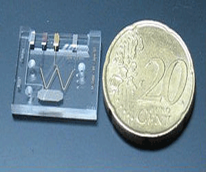
Actual size of a lab-on-a-chip
Thus, even sensors constituted by single molecules bound to their surface have been designed. Consequently the process of diagnosis is performed with a much smaller size device, improving portability and the potential for use anywhere.
Also, only extremely small sample quantities (micro or nanolitres) are required to carry out the analysis, so the sample extraction methods are less traumatic and invasive for patients.

These instruments also facilitate the use of samples without fluorescent or radioactive markers, which are used in biological and clinical analysis. This property is essential, since it has been proven that when a sample is not marked or altered before examining, the detection sensitivity significantly increases.

Within nanobiosensors we can distinguish the following categories:


Nanophotonic biosensors

This class of nanobiosensors, also known as evanescent wave biosensors, is based on how the light is transmitted by multiple internal reflections through the optical waveguide, which is a component of the biosensor.
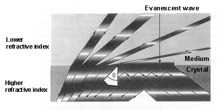
Evanescent wave productionWhen light is propagated in a medium and passes into a lower refractive index[1] medium, it is not totally reflected. As a consequence, a light component (called evanescent wave), in each of these reflections, is propagated in the lower refractive index that covers the waveguide. This propagation is really short, a few hundreds of nanometers, but it permits interaction between the light and the detection surface of the sensor, demonstrating which specific bioreceptors anchored to that surface interact with the analysed sample.

With these devices, only a few microliters of sample are necessary to determine protein concentrations or the variations of just one DNA base.

Also, it is possible to evaluate the metabolic state of just a single cell because some of these nanobiosensors have sharp fibre optic (30-50 nm) filaments, which can be introduced into cells through the plasma membrane without harming the cell.
This method facilitates the study of in vivo cell functions (apoptosis[2], cell division, biological nanomachines…) and it can detect pathological changes in a single cell.


Nanoplasmonic biosensors

In the Surface Plasmon Resonance (SPR) biosensor, a thin metal layer (normally a 50- nm-thick layer of gold) is placed on a dielectric material (a crystal), and its functioning is based on the detection of changes in the refractive index around the separation area between both elements.
By exciting the interface between these two layers (under conditions of total internal reflection) a plasmonic resonance[3] is activated with a certain angle of incidence.. This angle, which produces a plasmonic wave (with an evanescent character) is very sensitive to the changes from molecular interactions that happen on the metal surface. Therefore, these interactions between the analyte[4] of the sample and the sensor surface of the biosensor are shown as variations of the resonance angle.
Generation of Surface Plasmon Resonance (SPR)

Currently, new biosensors are being developed, which rely on the plasmon resonance phenomenon in nanoparticles. While their detection range would be very similar to the SPRs, their functioning system can be made simpler by measuring light transmission instead of light reflection, in addition to the advantage of the device miniaturisation.

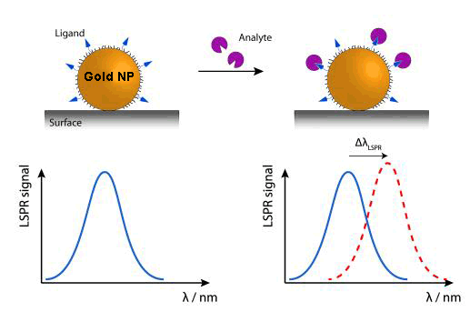
Due to the tiny size of nanoparticles, the electron oscillations are more localised than in the previous case (SPR), in specific areas of nanoparticles. This phenomenon is called ‘Localized Surface Plasmon Resonance’ (LSPR). The colour (wavelength: λ) changes that nanoparticles experience by adsorbing[5] the (bio)molecules of the examined sample are used to analyse this sample.
Variation of wavelength (Δλ) in nanoparticles when LSPR is produced

An alternative, within this kind of device,, is DNA sensors detecting colour changes produced by the aggregation of gold nanoparticles marked with stranded DNA complementary to the target DNA.

Nanoplasmonic biosensors are characterised by their real-time detection, specificity, high sensitivity and recognisition rate.
Their main applications are found in veterinarian, biomedical and environmental fields as well as in the food industry.


Nanomechanical biosensors

These sorts of nanobiosensors use the deflection or the resonance frequency variation of a microcantilever (as a transducing[6] method) when they interact with the studied sample. This position variation (Δx) is just a few nanometers derived from the biomolecular identification of the analysed sample.
Working principle of nanomechanical biosensors

Because these microcantilevers can be produced in mass at a low cost by standar microelectronic technology, thousands of them can be manufactured to identify thousands of analytes in a single sample.

The sensor area of these microcantilevers is around 1,000 μm2, which provides them to test sample volume lower than femtomole (10-15 moles).

Both photonic and nanomechanical biosensors could help us to obtain unlimited genetic and proteomic information that would offer the discovery of pathogens, new drugs, vaccines and undiscovered mutations of certain diseases in a faster way than the current technology.


Microfluidic devices or lab-on-a-chip (LOC)

This class of instruments uses an electric field to move liquids, particles, molecules or cells through microcapillaries designed on chips of different materials like silicon, crystal, quartz or plastic.
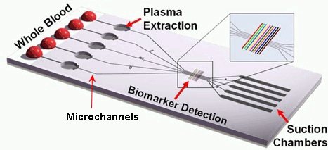
A schematic diagram of a  lab-on-a-chipThis kind of chip is configured by a large number of integrated microchannels and microchambers, where the examined sample is subjected to complex chemical and biochemical reactions.
The required volume of the sample fluids is very small, therefore the analysis can be carried out very quickly.
It is a portable and disposable detection method with a high grade of automation.

The ultimate objective for all these biocompatible nanodevices is to finally be implanted in vivo inside our organism, where they would perform a similar function to ‘sentinels’ in the presence of the first diseased cells. Some advances have already been made to that effect at the microscale (pills with built-in image cameras), but, without a doubt, this will become one the major research fields in nanomedicine in the near future.


[1] It describes how light propagates through a certain medium. It is defined as n = c/v, where c is the speed of light in vaccum and v the speed of light in that medium.
[2] Process of programmed cell death.
[3] Excitation or oscillation of conduction electrons at the interface between two materials, in our case a metal layer on a dielectric substrate.
[4] Substance (ion, compound or element) of interest in an analysed sample.
[5] Adhesion of atoms, ions or molecules to a surface.
[6] Process by one signal or energy is converted to another.

Sources: Nanomedicina: aplicación de la nanotecnología en la salud. Laura M. Lechuga. Grupo de Nanobiosensores y Aplicaciones Bioanalíticas
              Centro de Investigación en Nanociencia y Nanotecnología (CIN2). CSIC
              http://www.kennislink.nl/publicaties/de-opmars-van-de-twentse-lab-on-a-chip
              http://images.slideplayer.es/16/5040279/slides/slide_47.jpg
              http://www.mdpi.com/sensors/sensors-10-09630/article_deploy/html/images/sensors-10-09630f1-1024.png  
              https://www.ifm.liu.se/applphys/molphys/research/biosensing_using_nanopart/
              http://www.tcd.ie/Physics/people/Martin.Hegner/ReviewNSST-The_impact_of_STM_and_AFM.html?ntherodt_01.pdf
              Fritz, J., Baller, M.K., Lang, H.P., Rothuizen, H., Vettiger, P., Meyer, E., Guntherodt, H.-J., Gerber, CH. and Gimzewski, J.K., 
              Science 288 (2000) 316.
              https://www.theengineer.co.uk/lab-on-chip-device-promises-hiv-diagnosis-in-10-minutes/


Your opinion matters

3.4. Proteins

0 comments
Proteins are the biological macromolecules with the largest variety in structure and functions. We can find thousands of them in a single cell, each performing a unique and specific role. In this way, we find proteins carrying out transport or storage functions, protective, contractile and structural proteins, enzymes or toxins.
All of them have in common that they are made up of structural polymers of amino acids arranged in lineal chains.


Types and functions of proteins

      Enzymes: they are known as complex or conjugated proteins, whose essential function is to act as an organic catalyst in biochemical reactions, where they assist to synthesise, reorder or break down different elements on which they perform.
An example are digestive enzymes, like salivary amylase, hydrolyses amylose (starch component).
      Hormones: they control and regulate physiological processes such as metabolism, growth or reproduction. They are chemical-signalling molecules, usually stereoids or small proteins, released by endocrine cells.
Examples include insulin, which is a protein hormone in charge of regulating glucose levels in blood.

Proteins present a wide range of molecular weights and shapes, where shapes are vital to their activities. But these functions can be modified by the exposure to chemicals and changes in pH or temperature, which lead to shape variations, and as a consequence the loss of their functions in a process named denaturation.


Amino acids

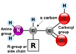
Amino acids are the monomers that constitute proteins. Each one has a structure formed by a central carbon atom, known as α carbon, to which an amino group (NH2), a hydrogen atom and a carboxyl group (COOH) are attached. The name ‘amino acid’ comes from the presence of these two groups in the amino acid basic structure.
General structure of amino acids
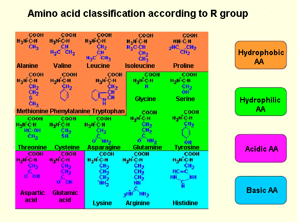
Each amino acid also have another atom or group of atoms joined to the central atom, called R group or side chain which is different for each kind of amino acid. The chemical nature of this chain establishes the type of aminoacid (whether it is polar, nonpolar, basic, acidic…). Thus, we can find basic aminoacids, like arginine and lysine, with a positive charge. Others such as cysteine or serine are polar due to their hydrophilic side chains, or if this chain is hydrophobic we have nonpolar amino acids like alanine or valine.
Amino acid classification according to R group
Out of the twenty different amino acids that form proteins, ten of them are considered essentials, because they are the necessary blocks for the protein construction. However, the human body cannot sinthesise them and must be obtained from the diet.

Amino acids are represented by a single upper case letter or three-letter abbreviation, e.g.: cysteine is symbolised by the letter C or the three letters Cys.

The number and sequence of amino acids ultimately set up the shape, function and size of proteins.

Amino acids are linked by covalent bonds, called a peptide bond, in which the carboxyl group of one amino acid is bonded to the amino group of the adjacent amino acid, releasing a water molecule during the process (dehydration reaction).
These bond products are called peptides and their joining give rise to a polypeptide chain. Each one has a free amino group at the end of the chain, named N terminal or amino terminal, having a C terminal or carboxyl terminal at the other end.

Often, the terms polypeptide and protein are used interchangeably, although technically a polypeptide is an amino acid polymer, while a protein is a polypeptide or combined polypeptides, each one with distinct shape and unique functions.


Protein structure

As I previously mentioned, protein shape is vital for the role they perform. In order to understand how a protein adopts its shape or final conformation, it is necessary to study the four structural levels in which they are organised: primary, secondary, tertiary and quaternary structures.

Primary structure
The primary structure is defined as the unique sequence of amino acids in a polypeptide chain. For example, the insulin hormone has two polypeptide chains, A and B, formed by unique sequences of 21 and 30 amino acids respectively and joined by two disulfide bonds between the cystein amino acids. There is also a third disulfide bond in the A chain, between two cysteine amino acids, which enables the molecule to fold into the suitable shape.

The unique sequence of every protein is ultimately determined by the genetic coding of that protein. Any alteration in the nucleotide[1] sequence, which constitutes the gene, can produce different amino acids that will be, later, added to the polypeptide chain and consequently affect the activity and structure of the resulting protein.

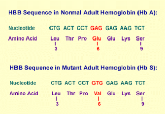
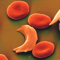
Thus, a single change in one of the 600 amino acids that make up the haemoglobin molecule origins, such a variation in its structure and activity, produces the appareance of sickle cell anaemia[2].Specifically, in the β chain of this protein, glutamic acid is substituted by valine.
More precisely, since every amino acid is made up of three nucleotide bases, those 600 amino acids generate 1,800 bases and sickle haemoglobin, the cause of sickle cell anaemia, arising from a single mutation in those 1,800 bases.
Mutant adult haemoglobin
Sickle cells
As a result of this, haemoglobin molecules form long fibres that deform the disc or biconcave shape of red blood cells, acquiring a sickle or crescent shape that blocks arteries. This obstruction causes a series of disorders such as headaches, abdominal pain, diziness or breathlessnes so typical of this sickness.

Secondary structure
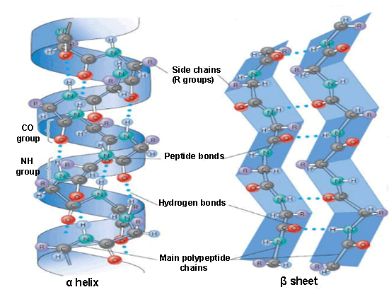
Secondary structures are generated from the local folding of polypeptides in certain regions, the most common are the α-helix and β-pleated sheet structures.
Secondary structures: α helix and β sheetThe α-helix is formed from the primary structure by rolling and twisting helically, with 3.6 amino acid residues per turn. They are constituted by hydrogen bonds between the carbonyl group (C=O) of one amino acid and the amino group (N-H) of another amino acid located four positions further in the polypeptide chain.

In the beta formation, amino acids are arranged in a zigzag way, named the ‘pleated sheet’ arrangement. Also it is originated by hydrogen bonds between N-H and C=O groups, but in this case they belong to adjacent chains.

Tertiary structure
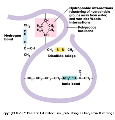
The tertiary structure informs how the secondary structure of a polypeptide folds up over itself giving rise to a globular conformation.Main bonds of tertiary structure
This configuration determines the physicochemical properties of proteins and assists their solubility in water, enabling, this way, it to carry out hormonal enzymatic and transport functions. This structure retains its stability due to the bonds among the R groups of amino acids. These bonds are created from the following types of interactions: disulfide bridges[3], hydrogen bonds, ionic and hydrophobic interactions and Van der Waals forces (to refresh all these concepts, visit the post titled: “Chemical bonds”).
In this arrangement, the nonpolar amino acids are, generally, facing the inside of the protein and the polar amino acids facing the outside, interacting with the surrounding aqueous medium.

Quaternary structure
In nature, some proteins are formed from several polypeptides, known as ‘subunits’, and their interaction creates the quaternary structure. These are weak interactions that assist the stabilisation of the overall structure of the protein.
The quaternary structure, usually, gives the protein its function and also creates many crucial biological structures, e.g.: viral capsids[4], microfilaments[5], microtubules[6] and collagen fibres of the connective tissue.
Structural levels of proteins


Denaturation and protein folding

Every protein has its own sequence and shape that are maintained through chemical interaction.

If the protein is exposed to chemicals or subjected to variations in pH or temperature, then its shape and structure can be modified without affecting its primary structure. This process is called denaturation, which can be reversible as long as the polypeptide primary structure is invariable during the process, allowing the protein to maintain its activity.
On the other hand, when the process is irreversible, the protein loses its function. This is what albumin (protein of egg white) experiences by being cooked, since it is denatured when it is exposed to high temperatures.

Protein folding plays a vital role for protein functions. This folding is made with the help of a protein family, named chaperones (or chaperonins), present in all cells. They are not part of functional proteins[6], but they bind to them to assist in their folding, their assembly and their cell transport to other parts of the cell where the protein performs its activity.


[1] Monomers of nucleic acids (DNA and RNA).
[2] Crescent or disc-shaped structures.
[3] Strong covalent bond between thiol groups (-SH) in the cysteine amino acid.
[4] Protein coat of virus that protects the viral genome.
[5] Thin fibres of proteins which with microtubules form the cell structure.
[6] Tubular structures of cells. Main component of cytoskeleton in eukaryotic cells.
[7] Those proteins that have a biological activity.

Sources: OpenStax College, Biology. OpenStax College. 30 May 2013.
              http://www.profesorenlinea.cl/Ciencias/ProteinasEstruct.htm
              http://study.com/academy/lesson/proteins-iv-higher-order-structure.html
              http://quimica.laguia2000.com/conceptos-basicos/cadena-lateral-en-aminoacidos
              http://es.slideshare.net/carolinacisdel/biomoleculas-enfermera
              http://biologiavfe.blogspot.com.es/2010/04/anemia-falciforme.html
              http://sicklecellcurefoundation.org/living-with-sickle-cell-disease/national-sickle-cell-month
              http://www.slideshare.net/VijayP7/secondary-structure-prediction-of-proteins
              http://www.slideshare.net/thelawofscience/protein-structure-11543259
              http://www.mobi4health.ug.edu.pl/wp-content/uploads/2014/10/2ndMSW-Milla-Neffling-Primer.pdf


Your opinion matters

1.3.5. Novel drug delivery systems

0 comments
Currently, there are two basic problems related to drug dosage.
The first when the drug is too soluble in water and a controlled release system is required to counteract this. The second when the drug, on the contrary, has no or very low solubility, and therefore, it does not dissolve in water.

This second problem is vital to solve because, today, it is estimated that between 75 and 80% of new chemical substances that come out of academic laboratories and pharmaceutical companies are not water-soluble.
This challenge is to get the main constituents of the drug to dissolve in the gastrointestinal tract, and, in this way, to perform their systematic action[1].

Thanks to nanoparticle engineering technologies, new crystalline drugs can be designed with a really small size (between 200 and 300 nm), which enables them to have faster dissolution rates than larger particles.

Once they are manufactured, these nanoparticles are incorporated into the different drug delivery systems where they are dissolved. In this way the drug is available to be absorbed by the lungs or the digestive system, depending on which route of administration has been chosen.

An alternative would be a drug Crystalline and amorphous structures dissolved in an organic solvent (not water) such as acetonitrile, methanol or ethanol. The drug is then dissolved with some kind of stabiliser in order to then be rapidly frozen. As a result of this procedure we obtain non-crystalline pharmaceutical substances, named amorphous forms or amorphous morphology, which can be dissolved at much higher concentrations than their corresponding (water) crystalline forms.
Thus ‘super-saturation’ of an aqueous solution is achieved, which facilitates the drug’s dissolution at high concentrations in the stomach and in the upper small intestine to be subsequently absorbed and distributed through the whole organism.

Properties of crystalline and amorphous drugs
Crystalline stateAmorphous state
Long range translational, rotational
and conformational order
May exhibit short range order
Well defined melting pointGlass transition point[2]
Good flow properties[3]Poor flow properties
More stableLess stable
Less hygroscopic[4]More hygroscopic
Relatively less solubleRelatively more soluble

An application example of this type of particle engineering system has been developed by members of the medical school of the University of Texas in San Antonio and the Veterans Administration hospital, looking at a new administration route for immune-suppressing drugs.

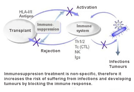
This sort of medication prevents the rejection of transplanted organs, but at present, by being administered orally it can cause a series of undesirable side effects, like the development of different types of cancer.
Side effects of immunosuppressant medications
The objective of the technologies described above was to administer a lower dosage of these drugs with accuracy to the site where its immune-suppressing action was necessary. In order to achieve this, prototypes of a new inhaled immune-suppressing drug were developed during a period of four to five years. The inhalation route of administration was selected because the patients who had undergone a lung transplant were the research target, and consequently the lungs were the aim of this medication.
Several tests were performed in mammals (mice and rats) where the product safety was proven. Low levels of the drug in the blood but high concentrations in the lungs were identified, which shows the validity of the inhalation route.
Then, the drug was tested in a small group of healthy human volunteers, under strict safety rules, and the results were similar to those in animals. Yet, this medication is still under development.

In conclusion, the goal is the research of new routes of drug administration that are not available presently to benefit the patients by minimising side effects and reducing the systemic drug exposure[5].


[1] Action exerted by the drug once is absorbed and distributed by the bloodstream.
[2] Temperature at which an amorphous solid becomes soft and flexible upon heating or rigid and brittle upon cooling.
[3] These properties indicate a higher or lower fluidity of particles depending on their size, shape, absorbed moisture and density.
[4] Substances that absorb moisture from the surrounding environment.
[5] A type of exposure where, in contrast with topical or local exposure drugs, the entire organism is affected by a medication performance.

Sources: UTAustinX: UT.4.01x Take Your Medicine - The Impact of Drug Development.
              http://www.slideshare.net/rcdreddi/limerick-meet-and-greet-ac-edited
              http://chemistry.about.com/od/chemistryglossary/a/glasstransition.htm
              https://docs.google.com/document/d/1TOwPCUSItCGOnUivKC_5kuTGw7kh9D5DPIK2U-V7D2A/edit#heading=h.90pmyxnddx1b
              http://trasplante-de-organos.blogspot.com.es/2011/09/inmunologia-del-trasplante-la.html


Your opinion matters

2.2.2. Imaging diagnostics

0 comments
Magnetic resonance scannerOver the last few years, imaging diagnostics has significantly increased its relevance such that they have become an indispensable tool to diagnose innumerable diseases such as neurological syndromes, cardiovascular disorders or cancer.

The convergence between imaging diagnostics and nanotechnology enables the creation of a new series of tracers and contrast agents with a higher resolution and sensitivity, which allows illness detection at earlier stages at cellular or even molecular level. This advance makes the quick application of treatment possible, thus increasing the chances of treatment and survival.

Imaging diagnostic nanosystems are mainly based on three types of nanoparticles: semiconductor, metal and magnetic.


Semiconductor nanoparticles

This first sort of nanoparticles, also called quantum dots, consist of semiconductors[1], whose size has been reduced to a few nanometres (1-10 nm). This produces changes in their electronic arrangement, which means they lose their distinctive band structure.
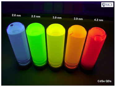
Because of this new structure, their optical response (specifically their fluorescence) is going to be closely related to the size variation of these nanoparticles. Consequently, these quantum dots emit light in a broad range of wavelengths (colours), depending on their size and not the material they are made of, which turns them into excellent biological markers.
Relationship between quantum dot size and wavelength emitted
While a wide variety of semiconductor materials have been examined, the most commonly used are cadmium selenide (CdSe) and cadmium telluride (CdTe), because in addition to be manufactured on a large scale, their sizes can be controlled perfectly. This enables us to see emission bandwiths with a full spectrum of different colours over a long lifetime.

But the semiconductor nanoparticles must also get to their defined targets. To achieve that, the quantum dots must be coated with polymers, like polyethylene glycol (PEG), which makes them invisible to macrophages and prevents immune system cells degrading and digesting them before reaching their target.

The next step is to make these nanoparticles able to identify their targets through bioreceptors (antibodies) that are bound to their surface, and these in turn will be bound to the specific antigens[2] of the target cells (e.g.: cancer cells). The bioreceptors of quantum dots produce a biomolecular recognition reaction when they meet these antigens, being accumulated in that area, and they are observed by using ultraviolet light due to the peculiar fluorescence that they emit.

Lastly and once they have fulfilled their role, the quantum dots must be purged from the organism to avoid unwanted side effects. It seems that the quantum dots are excreted through the kidneys and the liver without difficulties in animal testing, but to be tested in humans some problems related to the aggregation process must be resolved before receiving authorisation from the health agencies for commercialisation.


Metal nanoparticles

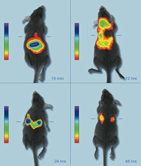
A live mouse model showing the accumulation of near infrared fluorescent nanoparticles in breast cancer tumorsThe fact that metal nanoparticles have a resonance frequency (their colour) dependent on their size and shape makes them a second option for contrast agents.
This property provides enables them to be manufactured to absorb or reflect light in the spectrum of interest. Thus, gold nanoparticles can be designed to absorb or reflect light in the near-infrared band (700-900 nm) as biological tissues are more transparent in that electromagnetic spectrum band. Using techniques like optical coherence tomography (OCT)[3], 3D maps are obtained of the areas where the nanoparticles are gathered.


Magnetic nanoparticles

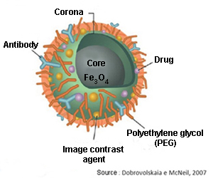
A third alternative is magnetic nanoparticles (iron oxides like magnetite: Fe3O4), with the ability to increase the contrast in magnetic resonance imaging (MRI) tests.
Functionalized magnetic nanoparticlesTheir transport through the organism can be carried out with the use of an external magnetic field (like an electromagnet) taking advantage of their magnetic properties. They may replace the old fashioned markers made of heavy metals in the near future, because they have lower toxicity.

Both magnetic and metal nanoparticles use identical or very similar methods to the ones described for the quantum dots in order to avoid degradation by the immune system, thus enabling them to locate and bind to the target tissues and cells.

Imaging nanosystems belong to the in vivo diagnostic category, therefore they must be injected into the human body. This implies, as we have seen, potential drawbacks (biocompatibility, sophisticated design) that have to be resolved to ensure an effective and safe use in the organism, minimising undesirable side effects.


[1] Material that behaves as an insulator or a conductive material depending on several factors such as environment temperature, pressure, surrounding electrical and magnetic fields or the incident radiation.
[2] External or toxic substances for the body (generally proteins) that lead to antibody production and causing an immune response. 
[3] Non-invasive imaging test that uses light waves to take cross section images.

Sources: Informe de vigilancia tecnológica: nanomedicina. Fundación para el conocimiento madri+d. CEIM. José Manuel González, Marta López, 
              Gema Ruiz.
              Nanomedicina: aplicación de la nanotecnología en la salud. Laura M. Lechuga. Grupo de Nanobiosensores y Aplicaciones Bioanalíticas
              Centro de Investigación en Nanociencia y Nanotecnología (CIN2). CSIC
              http://www1.radiology.ucsf.edu/research/labs/hyperpolarized-mri-tech-2/facilities_equipment
              http://www.osiconference.org/osi2015/presentations/Tu2.3%20Zahn.pdf
              http://www.nanowerk.com/news/newsid=7441.php (Image: Penn State)
              http://slideplayer.com.br/slide/74350/


Your opinion matters

2.2.1. Introduction to biosensors

0 comments
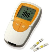
As a consequence of our current technology, the diagnosis of certain diseases is made at overly advanced stage of the disease.Example of biosensor: glucometer
Today, the main instrument used to diagnose illnesses is the biosensor, which enables the detection of specific substances, their composition and molecular features, or to identify the presence of microorganisms in certain environments.

This instrument is comprised of two main components:

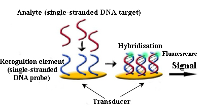
       A reactive or sensory area formed by a biological receptor (antibodies, DNA, enzimes…) with the ability to join those substances to be detected.
      A transducer system that processes and quantifies the signal (mechanical, electrical, optical…), which is caused by the interaction between the analysed substance and the recognition element.
The integration of both components makes biosensors sensitive and selective.
General scheme for biosensors
Biosensors have a wide range of applications in fields like environment, healthcare, biotechnology and the food industry.

Among the most common biosensors we find microarrays or biochips which consist of hundreds or thousands of biomolecules (generally oligonucleotides[1] or single-stranded DNA fragments, often known as ‘bioreceptor probes’) located on a solid surface (glass, silicon, gold) in particular positions and concentrations making a 2D matrix, with each molecule separated by a distance of between 100 and 150 μm from each other.
Biosensor for DNA detection
The examined sample is marked (normally with reactive fluorescent dyes), in such a way that, when it comes into direct contact with the sensory molecules of the biosensor, they are hybridised[2] with their homologous sequences, producing a fluorescent signal in that location.
Later, the data obtained are analysed and interpreted using a scanner and computer tools.

Biochips have countless applications: from personalised medicine (to know our higher or lower predisposition to suffer cancer or specific genetic disorders), the detection of harmful bacteria in food or water, discovery of virus and bacteria mutations, which become drug resistant or examining the microbial diversity in certain environments.

Another type of microarray are the protein microarrays (or protein chips) which, in contrast to DNA fragments, have thousands of functionally active proteins anchored to their surface. This class of instrument has great potential in basic research applied to molecular biology, the identification of disease markers and the search for therapeutic targets.

Lastly, we find within microarray technology cell chips. Antigens, proteins or even lipids are placed on these detector surfaces, which interact with cells, not only capturing them, but also triggering responses on them, such as phenotypic changes[3] or the segregation of particular substances.
These kinds of microarrays are used for toxicological analysis, identification of illness markers and the analysis of pathogenic agents.
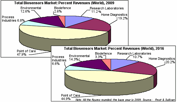
Evolution of total biosensors market: 2009-2016

Science keeps developing and it is designing smaller and more powerful devices, on the basis of the enormous potential that nanotechnology offers. These diagnostic nanosystems, whose main advantage is, in comparison to current biosensors, the early detection of diseases, depending on their working area, are divided into: imaging diagnostic nanosystems and nanobiosensors, which will be object of study in the coming posts within this section.


[1] General term for a short, single stranded DNA or RNA used in research, genetic testing and forensics.
[2] Recognition and combination of two complementary molecules
[3] Changes in those particular and genetically inherited features of an organism. 

Sources: Informe de vigilancia tecnológica: nanomedicina. Fundación para el conocimiento madri+d. CEIM
              Fundación española para la ciencia y la tecnología (FECYT). Nanociencia y nanotecnología. Entre la ciencia ficción del presente 
              y la tecnología del futuro, 2009.
              http://www.definicionabc.com/ciencia/fenotipo.php
              http://www.blogdefarmacia.com/biosensores-tecnologia-para-la-salud/
              http://www.pharmatutor.org/articles/applications-of-biosensors-technology-future-trends-development-and-new-intervation-in-                                   biotechnology?page=0,1
              http://www.slideshare.net/ManjuAnshika/biosensor-dr-manju-jha
              http://www.sensorsmag.com/specialty-markets/medical/strong-growth-predicted-biosensors-market-7640


Your opinion matters

1.3.4. Safety and efficacy of drugs

0 comments
In my second to last post (Routes of drug administration), we studied the different drug delivery systems and how one of them is selected to take a drug compound and make it into a drug product.

In the drug development process, researchers often use living organisms to find out if their compounds can go through the cell membranes and carry out their therapeutic function. However, there is a really delicate balance between those therapeutic effects and toxicity in lots of drugs. To avoid this conflict, previously, many studies must have been conducted to select the lead compound with the smallest toxicity and the top therapeutic potency.
Carrying out cell culture workMany of these evaluations can be assessed on the bench top in cell cultures. However, these experiments are often too simple to predict the compound efficacy in human beings. Hence, more complex living systems, like in rats or mice, are used in order to gain a greater understanding about their efficacy and biological distribution. To do this, researchers have been able to find several ways to model human diseases (e.g. cancer, certain infections and inflammations) in rodents.
Although these models are not completely reliable indicators of the drug efficacy in humans, they are another tool which helps to choose the lead compound with the greatest chance of success. These models can also be used to determine the optimal dosage of the drug needed to reach its target.

Despite the fact that in this phase, the pre-clinical efficacy is not considered as important as the pre-clinical safety, these studies are vital for the pharmaceutical developers to evaluate the likehood of the drug efficacy against a particular human disease. Once the lead compound has been selected and formulated, it is evaluated through animal testing to ensure it is safe for human use.
Animals used in research in the US
Although lots of ethical objections have arisen about animal testing, it is the best procedure that scientists and government officials have developed to protect humans against the unanticipated dangers of experimental drugs. In order to ensure the quality and validity of the obtained data in these trials, it has been set a strict group of regulations, called ‘good laboratory practice’ (GLP), which developers have to fulfil. These regulations establish an organisational framework for the new pharmaceutical substances, as well as the evaluation of any toxicity that may happen during examinations.

In addition to an understanding of the potential toxic effects, it is also critical to know how long the drug remains in the bloodstream, how it is metabolised and what tissues it can be collect in.
Format of IND

Next, I will explain how to obtain (on average) the permission of regulatory agencies to subject experimental drugs to clinical studies:
In the US, for instance, if the lead compound overcomes the safety exams, an IND (Investigational New Drug) application is submitted as evidence that the drug is reliable and that it can be put forward for experimentation in humans. Together with this data, the pharmacological product must be manufacturable by a reproducible and controlled method.

This stage in the drug development process, which can mean a financial layout of several million dollars for the pharmaceutical developers, can present an insurmountable obstacle for smaller companies and academic researchers.
In principle, the expenditure of such a huge amount of money on a new drug that has not been commercialised yet may appear excessive. Nevertheless, it is absolutely necessary to ensure that the drug will not cause harm to the humans that receive it during the clinical trials.

In the US, as I mentioned earlier, new drug products are given the denomination IND by the regulatory agency FDA (Food and Drug Administration), which does not mean that the new drug has been approved to be marketed, but the drug does then have authorisation to be put under clinical trial.
In order to get an IND, previously an IND application had to be submitted with all the required FDA information guaranteeing that the product is safe and ready to commence clinical trials.

During the first stage of clinical trials, the volunteers cannot be exposed, under any condition, to unnecessary health risks. To ensure this, the IND application must contain information about the safety test results of the drug, its manufacturing process and details about the proposed clinical study. As long as the provided information is clear and properly submitted, the FDA opens an IND application. In any case, the FDA could stop the investigation at any point if the study integrity is compromised or the new product turns out to be a health risk.
FDA organisation chart


Sources: UTAustinX: UT.4.01x Take Your Medicine - The Impact of Drug Development.
              https://www.msdsalud.es/tu-salud-al-dia/biblioteca-recursos/guias-para-pacientes/proceso-investigacion-desarrollo-aprobacion-
              farmaco.html
              http://www.thebody.com/content/art16845.html
              http://geneticayclonacion.blogspot.com.es/2015_04_01_archive.html
              http://speakingofresearch.com/facts/statistics/
              http://www.slideshare.net/swati2084/ind-investigational-new-drug-application-and-nda-26468953


Your opinion matters

1.3.3. A case study: developing inhaled products

0 comments
Delivering aerosols via inhalation has been practiced for millennia. As a matter of fact, since prehistory, where men began to use certain subtances with therapeutical and hallucinogenic purposes, such as cohoba (made from the beans of a mimosa species) or cannabis.

But aerosols only first replaced oral medication (tablets, pills, capsules…) in the 1950s for the treatment of respiratory diseases because oral medication caused severe side effects. The first meter-dose inhaler was made simply from a Coke bottle, some perfume valves and propellant systems.

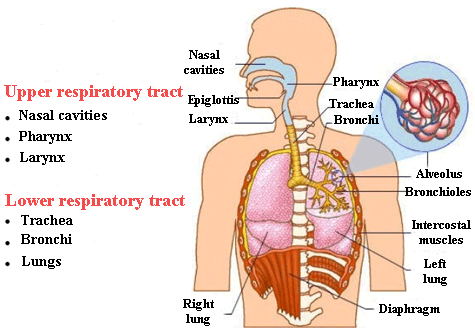
Respiratory system
The main challenge that aerosols face to penetrate the lungs and the upper airways (mouth, pharynx, larynx and trachea) is that these organs are very well equipped to filter dust and other particles. Hence, the challenge consists of designing the aerosol particles in such a way that they do not end up in the mouth or throat as opposed to the lungs.
These particles must be produced with a size or aerodynamic diameter between 1 to 5 μm, in order to pass beyond the upper airways, which act as a filter. As you can imagine, manufacturing such tiny particles is a hugely complex task.
For instance, those microscopic particles which are made of dry powder, have a very large surface area, which makes them very sticky. Consequently, the major force that acts on them is Van der Waals interaction (“Chemical bonds”) and not gravity (e.g. when you write with a chalk on a chalkboard it stays on and does not fall off due to these interactions rather than gravity). So, the objective is to overcome these Van der Waals forces and develop methods of deaggregating aerosol particles.

Lots of the aerosols used to treat pulmonary pathologies (e.g. cystic fibrosis, Chronic Obstructive Pulmonary Disease or COPD and asthma) are not really well designed for patient use.
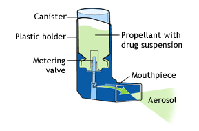
Metered-dose inhaler
For example, metre-dose inhalers have been used successfully for many years, however a lot of patients, especially children and the elderly, cannot or do not use them correctly. These inhalers present a major difficulty for those with coordination issues in activating the actuation manoeuvre with the inhalation manoeuvre.

On the other hand, the dry powder inhalers that we find currently on the market are allMultidose dry powder inhalers all active, which means that the patient needs to provide all the necessary energy so that the aerosol gets into the lungs via the airways.
The efficacy of these type of aerosols depends on how hard you inhale through the device. In this way, if you do not breath fast or strong enough, the aerosol particles will not be deaggregated enough to reach the lungs and they will mainly accumulate in the throat.
As a consequence, these sort of inhalers are not suitable for those patients who do not have proper lung function.
Portable nebulizer
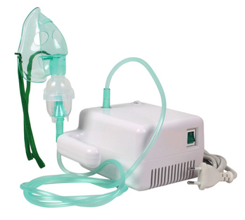
A third type of inhaler is a nebulizer or water-based nebulizer spray systems, usually bulky units, not very portable, used at home or in the clinic, although some technical developments have been made lately to make them more portable and battery operated.

So, these are the different devices for the treatment of respiratory diseases by the inhalation route and their selection is based upon the patient’s needs and the kind of drug to be delivered.

There is an interesting story about the formulation of the meter-dose inhalers. At the beginning, when these devices were invented, back in the 1950s, they used chlorofluorocarbon (CFCs) propellants into which the drug was suspended or dissolved. But, due to reports in the 1970s about ozone layer depletion because of the use of these propellant gases, and their subsequent ban in the late eighties, the pharmaceutical industry replaced them with hydrofluroalkanes (HFAs) with similar chemical and physical properties to the CFCs.

In spite of comparable properties, the HFAs behaved in a very different way with the pharmacological molecules and the rest of excipients. For example, some surfactants[1] used in drug formulation, like oleic acid, were soluble in the CFCs but not in the HFAs.

Because of this pharmaceutical companies adopted several strategies to reformulate the same drug into these propellants gases.
A meaningful example is the product called QVAR[2], in which the drug is dissolved, using a co-solvent (diluent), specifically ethanol, together with hydrofluroalkane propellants. And it turned out that the aerosol produced is a much finer mist with a much finer droplet size. In this way, the amount of aerosol particles deposited in mouth and throat decreased tremendously, but, on the other hand, there was a dramatic increase in lung delivery.
This new approach provided a competitive advantage for the company that created it in comparison to the competition, which tried to adapt the performance of the HFA propellants in a similar way to the older-style CFCs.


[1] Substance that act as a detergent, emulsifier or humectant and allows to reduce the surface tension in a fluid. 
[2] Anti-inflammatory inhaler indicated for the treatment of asthma attacks.

Sources: UTAustinX: UT.4.01x Take Your Medicine - The Impact of Drug Development.
              http://www.telegraph.co.uk/news/newstopics/howaboutthat/3225729/Stone-Age-man-took-drugs-say-scientists.html
              http://es.slideshare.net/CAWIMECA/el-sistema-respiratorio-42882484 
              http://blogs.20minutos.es/yaestaellistoquetodolosabe/tag/clorofluorocarbonos/
              http://definicion.de/surfactante/
              http://www.asthma.ca/adults/treatment/meteredDoseInhaler.php
              http://es.slideshare.net/luciagorreto/taller-asma-2015-inhaladores
              http://www.drtrust.in/products/nebuliser-machine


Your opinion matters

3.3. Lipids

0 comments
Lipids are a type of hydrocarbon mostly made up of nonpolar C-C and C-H bonds, which makes them hydrophobic (they repel water) or insoluble in water.
Lipids (more commonly known as fats) constitute a basic element in many hormones, as well as in cell membranes. They are an important source of long-term fuel for cells, they provide heat insulation for both animals and plants and they form a protective hydrophobic outer layer over fur or feathers of aquatic birds and mammals.
Within lipids we find the following categories:


Fats and oils

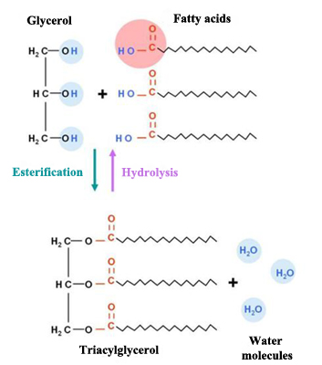
Fats, due to their chemical structure, are also named triacylglycerols or triglycerides. They are made up of two basic components: glycerol and fatty acids.
Glycerol is an organic component (specifically, a kind of alcohol) with three carbon atoms, five hydrogen atoms and three hydroxyl groups (OH). Fatty acids, in turn, are composed of a long chain of hydrocarbons to which a carboxyl group is attached.
The number of carbon atoms in fatty acids generally vary between four and thirty-six, although the most common contain 12 to 18 carbons. Fatty acids’ three carbon atoms (in the glycerol molecule) are joined by an ester bond and an oxygen atom. During the formation of this bond three water molecules are released.
Triglyceride formation
Fatty acids are divided into two groups: saturated and unsaturated.
The first only have simple bonds between the adjacent carbons of the hydrocarbon chain. An example is the stearic acid formed by 18 carbon atoms and in which the number of hydrogen atoms attached to the carbon backbone is the maximum (no more hydrogen atoms can be attached). This acid is present in oils and in animal and plant fats. Among its uses we find the manufacturing of soaps, candles, plastics, cosmetics and as a softening agent for rubber.

Unsaturated fatty acids, on the contrary, have double bonds between their carbon atoms. Among the most common unsaturated fatty acids we find oleic acid, known for its beneficial impact on blood vessels, thus decreasing the risk of liver and cardiovascular diseases.

Unsaturated fatty acids are popularly known as oils, which are liquid at room temperature and can be monounsaturated when they have one double carbon bond in the molecule (olive oil) or polyunsaturated if they have more than one (canola oil).
Adipose cells (adipocytes)

Plants store fats or oils in their seeds, which are used as energy deposits during their development. In mammals, however, fat constitute globules, which take up most of the volume of a very specialised kind of cell called an ‘adipocyte’.

Unsaturated fats or oils have usually a plant origin and contain cis unsaturated fatty acids (visit the entry “The carbon atom” to remember the concepts cis and trans configuration). This cis double bond causes a bend or “twist” which prevent fatty acids from being compacted, keeping them in liquid state at room temperature. As examples of unsaturated fats: olive, corn, canola and cod oils. These kinds of fats help us to reduce our cholesterol levels (in contrast to saturated fats which contribute to the formation of plaques in our arteries).

Trans fats
Trans fats are present in some food products like margarine or peanut butter and can lead to increased levels of low density lipoproteins (LDL), more commonly known as “bad” cholesterol, which is deposited on arteries walls as plaque, producing cardiovascular disorders.
These trans fats are manufactured by the food industry by a method called ‘hydrogenation’, in which the double bonds of the cis structure of the hydrocarbon chain become double bonds in the trans configuration. During this process, oils are solidified by injecting gaseous hydrogen into them, so that they can acquire that desirable semisolid consistency in many processed foods.

Omega fatty acids
Both omega-3 fatty acids and omega-6 fatty acids belong to the group of the essential fatty acids that our organism requires but is not able to synthesise; thus we must ingest them through our diet.
The terms omega-3 and omega-6 indicate that the third and the sixth carbon, counting from the far end of the hydrocarbon chain, are the ones that are attached to their adjacent carbon by a double bond.
Omega-3 fatty acidOmega-6 fatty acid


Among the food sources of omega-3 we find some oily fish like trout, tuna and salmon. These types of fatty acids reduce blood pressure and the triglyceride levels in blood, they help to prevent thrombosis, heart attacks and may help to decrease the risk of certain kinds of cancer.

Fats are not only excellent energy deposits in the long term and provide isolation for the body, but also enable us to digest fat-soluble vitamins. Therefore, despite the bad publicity that they have received, the “healthy” fats must be consumed in balanced diets regularly.


Waxes

Waxes are comprised long chains of fatty acids esterified[1] to long-chain alcohol.
Because of their hydrophobic nature, they perform a protective function on the outer coating of the leaves of some plants and the feathers of aquatic birds.
Esterification reaction


Phospholipids

Phospholipids are formed by fatty acid chains attached to a glycerol or sphingosine[2] backbone, where two fatty acids make up a diacylglycerol molecule and the third carbon of the glycerol backbone is occupied by a modified phosphate group.
The compound formed by the diacylglycerol molecule and the phosphate group constitute the phosphatidates, which are the precursors of phospholipids.
Phospholipids are part of the outer layer of animal cells and the main element of plasma membranes, which play a fundamental role in the cellular communication.
Phospholipid structure
Phospholipids make up these membranes in such a way that the phospholipid tails (which are hydrophobic fatty acids that cannot interact with water) face internally and the phospholipid head (which is the hydrophilic phosphate group that interacts with water) faces externally, in contact with the aqueous environment. By having a hydrophobic and a hydrophilic part, phospholipids are classified as amphiphatic molecules.
Phospholipid bilayers of cell membranesMicelle structure
The dynamic nature of plasma membranes is due to their being formed by phospholipids. In contact with water, phospholipid molecules are ordered spontaneously in a spherical structure called a micelle, with the heads (polar) facing the outside and the tails (nonpolar) facing the inside of these structures, just like plasma membranes.


Steroides

In spite of the fact that steroids are not very similar to the rest of the lipids, on account of the fact that they have a fused ring structure, they are included within this category as they are insoluble in water.
All steroids are made up of four carbon rings and many of them also have the –OH functional group, which allows them to be classified as alcohols (sterols).
In addition, some of them, like the cholesterol molecule, have a short hydrocarbon tail. Cholesterol is the most common steroid in the human being and animals, it is synthesised in the liver and is the precursor to vitamin D and also of bile salts, which help to metabolise the fats we ingest to be absorbed by cells afterwards.
Cholesterol is secreted by the endocrine glands and the gonads, as it is the precursor of steroid hormones like estradiol[3] and testosterone. Therefore, despite the bad name that cholesterol has among lay people, this molecule plays a vital role for the proper functioning of our organism.
Schematic classification of lipids


[1] Esterification reactions are those by an ester is obtained, usually from the reaction of a carboxylic acid and an alcohol.
Esters are organic compounds made by substituting an acid by an alkyl or other organic group.
[2] Amino alcohol made up of 18 carbons, forming an unsaturated hydrocarbon chain.
[3] Female sex hormone.

Sources: OpenStax College, Biology. OpenStax College. 30 May 2013.
              http://www.genomasur.com/BCH/BCH_libro/capitulo_02.htm
              http://www.salud180.com/sustancias/acido-estearico
              http://herbolaria.wikia.com/wiki/%C3%81cido_oleico
              https://www.flickr.com/photos/thame/3302072732
              http://www.eufic.org/article/es/artid/La-importancia-de-los-acidos-grasos-omega-3-y-omega-6/
              http://www.uhu.es/08007/documentos%20de%20texto/apuntes/2005/pdf/tema_03_lipidos.pdf
              http://www.fisicanet.com.ar/biologia/introduccion_biologia/ap11_lipidos.php
              http://brainly.com.br/tarefa/453268
              http://www.calpoly.edu/~jfernsle/Research/Biophysics/BiophysResearch.html


Your opinion matters

1.3.2. Routes of drug administration

0 comments
After gathering all possible information about the lead compound, researchers must now understand how to manufacture a drug product. This applies not only to the lead compound but also to other chemicals that favour its disolution in the gastrointestinal (GI) tract, help in the manufacturing process or protect it from degradation.

In addition to all these subtances, a wide array of products are added. In tablets, for example, cellulose is often included to improve the drug disintegration in the digestive fluids. Talc is also included in low concentrations during the manufacturing to absorb liquid and semi-liquid subtances. Lactose, or milk sugar, is often aggregated to bulk the tablet, acquiring a convenient size so that the patient can hold and swallow it.
Some tablets contain functional polymers to develop certain drug attributes. One of the main uses of functional polymers is to protect the tablet from the high acidity in the stomach, thus ensuring that the drug is dissolved in the small intestine later, and in this way improving its absortion in the intestinal fluids.

Even though a drug can be administered in many different ways, I will focus on those drugs that are administered orally to focus on the following questions:
Will the drug be dissolved and absorbed?, depending on how much it is absorbed, what should the dose be?, is it metabolised by the liver quickly after its absortion?, will it be degraded by the acidic media in the stomach?
With regard to the last question many drugs can be degraded at low pH.
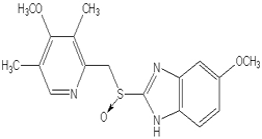
Chemical structure of Omeprazole
An example of these kinds of drugs is omeprazole, which without a functional polymer would not be effective. The purpose of this polymer is to preserve the omeprazole granules from direct contact with the gastrointestinal fluid until the pH becomes less acid and the drug is no longer in danger of being degraded. For this reason, it is important to remember that tablets and pills must not be broken, unless the pharmacist indicates it, or the polymer coating would be damaged and the drug would not be effective.
Routes of drug administration
Undoubtedly, the most usual way of delivering drugs to the body is orally because pills, capsules and tablets are easy to administer, mass-produced, have good stability and are easily absorbed by the digestive system.
But there are other systems that, at times, are better alternatives to the oral medication. Thus, intravenous or IV administration is performed when a high or well-controlled dose is required. This is the case with highly toxic agents (chemotherapy). However, this system has as a main disadvantage that is an invasive technique and it requires a clinician as well as sterile material to be dispensed.
Topical administration is another delivery system, for example ointments and creams to relieve irritations, rashes or insect bites as well as transdermal patches and gels which can permeate the skin so that the drug reaches the bloodstream.
Major routes of drug administration
A fourth type are inhaled medications, which like the former, perform locally, helping to treat illnesses like asthma, allergies or chronic lung diseases.Taking advantage of the fact that lungs are covered with a vast amount of vessels and capillaries, lots of drugs are inhaled (whether they are specific for the treatment of lung diseases or not) with the goal of being rapidly taken up by the circulatory system. As an example we can mention loxapine; an antipsychotic used in the treatment of schizophrenia, which when it is inhaled its effects can be detected in just ten minutes.

Therefore, the election of the suitable route of drug administration can make easier the drug uptake or even avoid possible side effects.


Sources: UTAustinX: UT.4.01x Take Your Medicine - The Impact of Drug Development.
              http://generalidadesdelafarmacia.blogspot.com.es/2010/11/vias-de-administracion-de-medicamentos.html
              http://www.xatakaciencia.com/medicina/farmacologia-vias-de-administracion-de-los-farmacos-y-sus-pros-contras              
              http://www.riesgoquimico.es/2009/03/30/omeprazol/


Your opinion matters